Sign in

Libre University uses your GitHub account. Signing in is only needed to sit a final test, so the score is kept on your profile.

The Cell

The machinery every living thing shares: membranes and transport, metabolism, the flow from DNA to protein, and how a cell divides.

What a cell is

Everything alive is built from cells, and the interesting part of that sentence is that it is a discovery rather than a definition.

This subject assumes nothing beyond ordinary school science: that matter is made of atoms, that a chemical reaction rearranges them, and that energy is conserved. Everything else is built here. Where a piece of chemistry or physics is needed, it is derived at the point of use rather than assumed.

Three claims, each of which had to be earned

Robert Hooke put a shaving of cork under a compound microscope in 1665 and saw a honeycomb of empty boxes. He called them cells, after the small rooms of a monastery, and he counted them: sixty across a span of one eighteenth of an inch, which makes each box a thousand and eightieth of an inch across, which makes a cubic inch of cork contain 10803=1{,}259{,}712{,}000 of them. He was looking at the dead cell walls of a plant, not at anything living, but the number is the first honest measurement in the subject.

Antonie van Leeuwenhoek, grinding single lenses in Delft, went further in the 1670s and saw things that moved. His letters to the Royal Society describe bacteria from his own dental plaque, protists in pond water, and sperm cells. Nobody could reproduce his lenses for a century, which is why his observations were doubted, and it is worth noticing that the first person to see a living cell was believed largely on the strength of his reputation for care.

The generalisation took another one hundred and sixty years and arrived in three parts. Matthias Schleiden argued in 1838 that all plant tissue is cellular, and Theodor Schwann extended it to animals in 1839, which is the claim that everything alive is made of cells. Schwann then got the second part badly wrong: he thought cells crystallised out of a formless fluid. Rudolf Virchow supplied the correction in 1855 with the slogan omnis cellula e cellula, every cell from a cell, and Louis Pasteur turned it from a slogan into an experiment. His swan-necked flasks, reported from 1859, held sterile broth open to the air indefinitely, because dust settled in the bend of the neck and never reached the liquid. Tip the flask so that the broth touched the dust and it clouded within a day.

So the cell theory is three separate empirical claims: living things are made of cells, the cell is the smallest unit that is itself alive, and cells arise only by the division of existing cells. None follows from the others. The third is the one that matters most for this course, because it means that the machinery inside a cell is never built from scratch. It is always inherited, running, from a cell that was already running.

Why life is packaged at all

A cell is a bag, and it is worth asking what the bag buys. The answer is that every living thing maintains a chemical composition different from its surroundings, and a difference in composition is only meaningful if there is a boundary across which it exists.

The numbers are not subtle. Human blood plasma carries about 5 mM potassium and 145 mM sodium. The inside of a human cell runs the ratio backwards, roughly 140 mM potassium and 12 mM sodium. Seawater is about 10 mM potassium. A cell is not in equilibrium with anything, and a system that is not in equilibrium with its surroundings is either being held there or is relaxing towards it. Life is the first case, permanently, which means it costs something permanently.

That is the whole architecture of the subject in one sentence. A boundary makes the difference possible, machinery in the boundary maintains it, and the maintenance has to be paid for. The next five lessons are the boundary and the payment.

The physics that sets the size

Nothing so far says how big a cell can be. That is set by diffusion, and diffusion has a property that is easy to state and easy to underestimate: the time it takes grows as the square of the distance.

A molecule in solution is knocked about by collisions and performs a random walk. The mean square displacement after time t in one dimension is x2=2Dt, where D is the diffusion coefficient, so the time to cover a distance x is

t=x22D

For a small metabolite in water D is about 5×10⁻¹⁰ m²/s, and cytoplasm is a few times slower than water because it is crowded. Put numbers in. Over 1 µm, the length of a bacterium, t=(10-6)2/(2×5×10-10)=10-3 s, one millisecond. Over 100 µm, a large animal cell, it is 10 s. Over 1 mm it is 1000 s, about seventeen minutes. Over 1 m it is 109 s, which is thirty-two years.

That is the constraint. A bacterium can run its entire chemistry on diffusion alone, with no transport system at all, because everything inside it meets everything else a thousand times a second. A millimetre of undifferentiated tissue cannot. The quadratic term means the penalty for growing is not proportional but brutal: ten times the radius is a hundred times the mixing time, while the volume that has to be supplied has gone up a thousandfold and the surface available to supply it only a hundredfold. The ratio of surface to volume for a sphere is 3/r, so a cell of radius 1 µm has ten times the membrane per unit of contents that a cell of radius 10 µm has.

Example. A ball of cells 1 mm across grows in culture with no blood supply. Using the diffusion law above, argue from the numbers whether its centre can be fed, and say what you would expect to find there.

The centre is 0.5 mm from the surface, so a small nutrient molecule needs t=(5×10⁻⁴)2/(2×5×10-10)=250 s to arrive, about four minutes. That does not sound fatal, and taken alone it is not: diffusion delivers, slowly. The killer is that every cell on the way consumes as the molecule passes, so the concentration falls with depth and the supply reaching the centre is what is left over. Real tumour spheroids show exactly this: a proliferating outer shell about 100 to 200 µm thick, a quiescent layer beneath it, and a necrotic core. The threshold sits near the distance oxygen can travel while being consumed, which is why capillaries in tissue are spaced tens of micrometres apart rather than millimetres. The general lesson is that diffusion sets a supply radius, and any organism that wants to be bigger than that radius has to invent plumbing.

Now you. A motor neuron in your spinal cord has an axon reaching a metre to your foot. The diffusion law says a protein made in the cell body would take decades to arrive. What must be true for such a cell to work at all?

Answer

Two things, and both are real. First, the cell cannot rely on diffusion for delivery, so it must move material actively: motor proteins walk cargo down microtubule tracks at roughly 1 µm/s, which covers a metre in about twelve days rather than thirty years, and that is exactly what axonal transport is. The eventual mechanism, kinesin taking eight nanometre steps for one ATP each, is the subject of a later lesson. Second, the cell cannot signal by diffusion either, which is why a nerve impulse is not a chemical travelling down the axon but a voltage change propagating at up to 100 m/s, using ion gradients the next two lessons build. The neuron is not an exception to the diffusion limit. It is an object built entirely out of ways around it.

One bacterium, counted

Take Escherichia coli, the best-quantified organism on the planet. A cell is a rod about 2 µm long and 0.8 µm across, so its volume is π(0.4)2×2=1.0 µm³, which is 10-15 litres, one femtolitre. Roughly seventy per cent of that is water.

Inside it are about three million protein molecules of some four thousand different kinds, with a mean mass near 40 kDa. Check that against the volume: 3×106×40{,}000 daltons, at 1.66×10⁻²⁴ g per dalton, is 2.0×10⁻¹³ g in 10-15 litres, which is 200 g per litre. That is a strikingly high figure. Cytoplasm is not a dilute solution, it is closer in consistency to a thick syrup, and the crowding is dense enough to change reaction rates and to be one reason diffusion inside a cell is slower than in water.

The genome is a single circular DNA molecule of 4,641,652 base pairs in the standard K-12 strain. At 0.34 nm per base pair that is 4.64×106×0.34 nm =1.58 mm of DNA, packed into a cell 2 µm long, a ratio of about 790 to 1. A human cell is worse: 6.4 billion base pairs is 2.2 m of DNA folded into a nucleus around 6 µm across. Any account of how DNA works has to explain how a molecule that long is stored, and how a polymerase finds one specific spot on it.

Example. An E. coli cell in rich medium divides every 20 minutes. Using the three million proteins above, work out how fast it builds protein, and then estimate how many ribosomes that requires if each one adds twenty amino acids per second.

To double, the cell must make three million proteins in 1200 s, which is 3×10⁶/1200=2500 complete proteins per second. At a mean length of 300 amino acids that is 7.5×10⁵ peptide bonds per second. A ribosome working flat out at twenty residues per second therefore supports 7.5×10⁵/20=37{,}500 of them, so the cell needs something like forty thousand ribosomes. Measured counts for fast-growing E. coli run from about 45,000 to 70,000, so an estimate built from two textbook numbers lands within a factor of two of the real thing. That agreement is worth more than either number alone: it says the picture is roughly right, and it says translation is not a minor activity of a cell but its single largest construction project.

Now you. The same cell contains about 4,000 kinds of protein but only one copy of its genome. What does that force to be true about how genes are used?

Answer

A single template has to serve every copy, so the gene cannot be consumed by being read. Reading must be non-destructive and repeatable, which rules out any scheme where the gene is converted into the product. It also means the gene is a bottleneck: with three million protein molecules and one copy of each gene, the average gene has been read out into hundreds or thousands of products, so there must be an amplifying intermediate between the gene and the protein rather than a one-to-one correspondence. That intermediate is messenger RNA, and the fact that its existence can be argued for from a counting argument, before any experiment, is a good sign that the eventual experiment was looking for something real.

Two ways of building a cell, and one thing they share

Cells come in two architectures. A bacterial cell has its DNA loose in the cytoplasm and no internal membranes to speak of. A eukaryotic cell keeps its DNA inside a nucleus and is filled with membrane-bounded compartments: mitochondria, endoplasmic reticulum, Golgi, lysosomes. It is also far larger. A typical animal cell of 15 µm diameter has a volume near 1800 µm³, roughly two thousand times a bacterium, and a plant or fungal cell larger still.

The old division into prokaryote and eukaryote hides something important. Carl Woese, comparing ribosomal RNA sequences in 1977, found that the organisms lumped together as bacteria are two groups as different from each other as either is from us. Bacteria and archaea look alike under a microscope and are not close relatives. "Prokaryote" is therefore a statement about what a cell lacks, which is a poor basis for a category, and it survives because it is useful shorthand rather than because it names a natural group.

What all three domains share is the striking part, and it is the reason this subject exists. Every cell yet examined is bounded by a lipid bilayer, stores its inheritance as double-stranded DNA, reads it into RNA and then into protein by ribosomes built of RNA, uses very nearly the same genetic code, and pays for its chemistry with adenosine triphosphate. That common core is why one course can be about "the cell" rather than about a catalogue of organisms.

Where the theory frays

An honest course names the exceptions early. A mature human red blood cell has ejected its nucleus and contains no DNA at all, so it cannot divide and cannot be the origin of another cell; it is a cell by descent rather than by capacity. A skeletal muscle fibre is a single cell up to 30 cm long containing hundreds of nuclei, formed by cells fusing, which makes the boundary of "one cell" a matter of definition. Fungal hyphae are often similarly continuous. Thiomargarita magnifica, described in 2022, is a bacterium up to 2 cm long, visible without a microscope, which manages the diffusion problem by confining almost all its volume to a fluid-filled vacuole so that the living layer stays thin.

Viruses fail the theory outright. A virus has a genome and a protein coat, no membrane of its own in many cases, no ribosomes, no metabolism, and reproduces only by commandeering a cell. Whether it is alive is a question about the word rather than about the virus, and the useful version of the question is what the minimum equipment for autonomous life turns out to be: a boundary, a genome, ribosomes, and a way of making ATP. That list is the syllabus of this course.

Example. A colleague says the cell theory is circular, since a cell is defined as the unit of life and life is defined as being made of cells. Is the objection fair?

Not really, and seeing why is worth the effort. The theory makes claims that could have come out false and did not. It could have turned out that tissue is a continuous substance in which nuclei are scattered, which is what the reticular theory of the nervous system asserted until Cajal's staining showed neurons to be separate cells. It could have turned out that cells form spontaneously from broth, which is what Schwann believed and Pasteur disproved with an experiment anyone can repeat. It could have turned out that different tissues are built on different principles. Each is a real alternative that was defeated by evidence, which is the mark of a claim with content. The definitional element is real but small: the boundary cases above show that "one cell" sometimes has to be stipulated. Stipulating a boundary at the edges is not the same as assuming the answer in the middle.

Now you. A giant amoeba and an ostrich egg are both single cells far larger than the diffusion limit suggests. Before reading on, say what you would look for in each to explain how they manage.

Answer

Look for either active stirring or an inert bulk. Large amoebae and plant cells take the first route: cytoplasmic streaming, driven by motor proteins along actin filaments, physically circulates the contents so that transport is bulk flow rather than diffusion, and a slow current across a millimetre beats diffusion easily. An ostrich egg takes the second: most of its volume is stored yolk, chemically inert, and the living cytoplasm and nucleus occupy a small disc a few millimetres across on its surface. The same trick appears in Thiomargarita, where a vacuole fills the middle. The general rule is that no cell escapes the diffusion law, so a large one either pumps its contents around or arranges for most of its volume not to be alive.

Everything in this lesson depended on there being an inside and an outside, and nothing in it said what the boundary is made of. That turns out to be answerable with an experiment from 1925 involving red blood cells, acetone and a trough of water, and the answer sets up the central difficulty of the next several lessons: the boundary that makes a cell possible is almost perfectly impermeable to the things a cell needs.
The membrane

The boundary that makes a cell possible was measured before anyone had seen it, using red blood cells, acetone and a shallow trough of water.

The previous lesson ended with the claim that a cell holds a composition different from its surroundings and must therefore have a boundary. This lesson asks what the boundary is made of, how thick it is, and how well it works. The last question turns out to matter most, because the answer is "far too well".

Why oil and water settle the question before any experiment

Charles Ernest Overton, working in Zurich in the 1890s, tested thousands of compounds for how readily they entered plant cells and found one predictor that beat everything else: how well the compound dissolved in olive oil. Substances that partition into oil crossed easily, substances that stay in water did not, and this held regardless of molecular size within the range he tested. Overton concluded in 1899 that the cell boundary is made of something oily, probably a lipid, and he was right thirty years before there was any direct evidence.

The reason a lipid boundary forms at all is not that lipids attract each other, which is the intuitive but wrong explanation. It is that water is strongly self-attracting, through hydrogen bonds, and a hydrocarbon chain sitting in water forces the surrounding molecules into an ordered cage that costs entropy. Push the hydrocarbons together and that ordered water is released. The driving force is the freed water, not the oil, which is why the effect is called the hydrophobic effect and why it gets stronger, not weaker, as temperature rises over the ordinary range.

The size of the effect can be read off a measurement. Phospholipids in water aggregate above a critical concentration of roughly 10-10 M, and the free energy of moving one lipid from solution into an aggregate is ΔG=RTln(CMC), which at 310 K is 2.577×ln(10-10)=-59 kJ/mol. Compare a single-chain detergent such as sodium dodecyl sulfate, whose critical concentration is about 8 mM, giving 2.577×ln(0.008)=-12 kJ/mol. Two tails instead of one is worth roughly five times the binding energy, and that factor is the whole difference between a soap, which forms small micelles and dissolves membranes, and a phospholipid, which forms sheets and makes cells. At -59 kJ/mol a lipid essentially never leaves the membrane it is in. The structure is not held together by anything; it is held together by water refusing to accommodate it.

Geometry decides sheet rather than sphere. A single-tailed detergent is a wedge, wide head and narrow tail, and wedges pack into a small sphere. A two-tailed phospholipid is closer to a cylinder, and cylinders pack into a flat sheet. A sheet has edges where hydrocarbon meets water, which is expensive, so a large enough sheet closes on itself into a sealed vesicle. A cell membrane is therefore self-assembling, self-sealing and self-repairing, none of which had to be designed.

Two molecules thick, measured in 1925

Evert Gorter and François Grendel, in Leiden in 1925, did the experiment that fixed the thickness. They took red blood cells from several mammals, counted them, extracted their lipids with acetone, and spread the extract as a single layer on the surface of water in a Langmuir trough, where a movable barrier compresses the film until it just becomes continuous. Reading the area of the film and dividing by the total surface area of the cells they had started with, they got a ratio close to two, and concluded that the membrane is a lipid layer two molecules thick.

The conclusion is correct and the experiment was flawed twice over. Acetone does not extract all membrane lipid, so their film was too small by roughly a third. And they estimated a red cell's surface area at about 99 µm², whereas the biconcave disc actually has about 145 µm², so their denominator was too small as well. Correct only the area and the ratio falls to 2×99/145=1.37, which would have suggested something other than a bilayer. Correct the extraction too, dividing the film area by 0.7, and it returns to 1.37/0.7=1.95. Two compensating errors gave the right answer, which is a good reason to be suspicious of clean results and a good reason not to dismiss them either.

The modern picture agrees. The hydrocarbon core of a bilayer is about 3 nm thick and the whole structure with head groups is 4 to 5 nm, so a membrane is roughly one thousandth the width of the cell it encloses. Each lipid occupies about 0.65 nm² of surface, so a red cell with 140 µm² of membrane and two leaflets carries 2×1.4×108/0.65=4.3×108 lipid molecules, and the measured figure is close to that.

Example. Suppose Gorter and Grendel had used a modern lipid extraction, recovering everything, but had kept their 99 µm² figure for the cell area. What ratio would they have found, and what would they have concluded?

Their film area would have been 198/0.7=283 µm² per cell, and dividing by the assumed 99 µm² gives 2.86. A ratio approaching three invites the conclusion that the membrane is three lipid layers thick, which is not a structure that makes chemical sense, since a middle layer would have to bury its head groups in hydrocarbon or its tails in water. The instructive part is what a careful reader should do with a puzzling ratio: check the denominator first. Cell surface area is the hardest quantity in the experiment to measure and the easiest to get wrong, because it depends on knowing the cell's shape rather than just its volume, and a biconcave disc has far more surface than the sphere of the same volume. The lipid film area, by contrast, is read directly off a calibrated trough.

Now you. Bacterial membranes contain no cholesterol, while an animal plasma membrane may be up to forty per cent cholesterol by molecule. Given that cholesterol is a rigid flat ring system with a single small hydroxyl group, what would you predict it does when inserted between phospholipids?

Answer

It should sit with its hydroxyl at the head group level and its rigid rings alongside the upper part of the fatty acid chains, and it should have two effects that sound contradictory and are not. Below the melting temperature of the lipids it gets in the way of orderly packing, so it keeps the membrane from freezing into a rigid gel; above it, the rigid ring restrains chain motion, so it stiffens a membrane that would otherwise be too fluid. Cholesterol therefore buffers fluidity against temperature rather than raising or lowering it, which is what a cell held at a fixed 37 degrees Celsius needs less than a cell that experiences weather. It also plugs the transient gaps between chains, and measured permeability to water and small solutes falls substantially when cholesterol is added. Bacteria solve the same problem differently, with hopanoids or by adjusting how many of their fatty acid chains are unsaturated, which is a real example of two lineages converging on a requirement rather than on a molecule.

A mosaic, and a fluid one

For thirty years after Gorter and Grendel the standing model was Danielli and Davson's 1935 sandwich: a lipid bilayer coated on both faces with sheets of protein. Electron microscopy in the late 1950s appeared to confirm it, since a thin section stained with osmium showed two dark lines with a light one between, and J. David Robertson generalised this "unit membrane" to every membrane in the cell.

It was wrong, and two lines of evidence killed it. Membrane proteins, once anyone managed to purify them, turned out to be largely hydrophobic on the outside, which is the wrong surface chemistry for something that lies on a membrane rather than in it. And freeze-fracture electron microscopy, which splits a frozen membrane between its two leaflets, showed the interior studded with particles 6 to 10 nm across, exactly what proteins crossing the bilayer would look like from inside.

The fluidity was demonstrated by Larry Frye and Michael Edidin in 1970 with an experiment of unusual clarity. They fused a mouse cell to a human cell to make a single hybrid, then labelled mouse surface proteins with an antibody carrying one fluorescent dye and human surface proteins with another. Immediately after fusion the hybrid was half red and half green. Within forty minutes at 37 degrees Celsius the two colours were completely intermixed over the whole surface. Nothing had been synthesised or destroyed in that time; the proteins had simply diffused.

Seymour Singer and Garth Nicolson assembled this into the fluid mosaic model in 1972: a two-dimensional lipid solvent in which proteins float, some spanning the bilayer, some anchored to one face. The lateral diffusion coefficient of a lipid is about 1 µm²/s, so with t=x2/4D in two dimensions a lipid crosses a 2 µm bacterium in (2×10⁻⁶)2/(4×10-12)=1 s. Flipping from one leaflet to the other is a different matter entirely, because it requires dragging a polar head group through the hydrocarbon core, and spontaneous flip-flop takes hours to days. That asymmetry is the reason the two leaflets can have different compositions and keep them.

They do. Phosphatidylserine, which carries a negative charge, is held almost entirely on the cytoplasmic face by dedicated flippase enzymes. When a cell dies by apoptosis the flippases stop and a scramblase runs, phosphatidylserine appears on the outer face, and macrophages recognise it and eat the cell. A lipid on the wrong side of a 5 nm sheet is a death certificate, which is a useful reminder that the membrane is not only a container.

The mosaic is dense. Protein makes up about half the mass of a typical plasma membrane, rises to 76 per cent in the inner mitochondrial membrane, which is a machine rather than a wall, and falls to 18 per cent in myelin, which is insulation and wants to be nothing but lipid.

Making more of it

A dividing cell has to double its membrane, and it cannot do so the way it doubles a protein. Lipids are synthesised by enzymes embedded in a membrane that already exists, using glycerol phosphate and activated fatty acids, and the product is inserted into the leaflet the enzyme faces. No cell builds a bilayer from nothing. Every membrane in every cell is an enlarged piece of a membrane that was there before, which is Virchow's principle from the first lesson applied to a structure rather than to a cell.

Two consequences follow. Because synthesis happens on one face, a flippase must move roughly half the new lipid across to keep the two leaflets growing together, which is the same machinery that maintains the asymmetry described above. And because a eukaryotic cell makes almost all its lipid in the endoplasmic reticulum, that lipid has to be distributed to every other compartment, which happens both by vesicles and at close contact sites where two membranes come within about 20 nm and lipids are handed across by transfer proteins.

Example. An E. coli cell is a rod 2 µm long and 0.8 µm across, and doubles every 20 minutes. Estimate how many lipid molecules per second its inner membrane requires.

The surface area is the cylinder plus the two caps, π×0.8×2+2π(0.4)2=6.0 µm², which is 6.0×10⁶ nm². At 0.65 nm² per lipid in each of two leaflets that is 2×6.0×106/0.65=1.9×107 lipids. Doubling in 1200 s therefore requires about 1.9×10⁷/1200=15{,}000 lipid molecules a second, and a gram-negative bacterium with a second, outer membrane needs roughly twice that. The number is worth having because it puts membrane synthesis on the same footing as the protein synthesis rate derived in the previous lesson, around 2500 proteins a second: lipid assembly is a major continuous manufacturing activity rather than a trickle, and a cell devotes a serious fraction of its acetyl-CoA to it.

Now you. Some antibiotics, and the antifungal drugs of the azole class, target lipid synthesis rather than protein or DNA synthesis. Given the numbers above, why is that a reasonable target, and what determines whether such a drug can be selective?

Answer

It is reasonable because membrane synthesis is continuous, fast and essential: a cell that cannot make lipid cannot grow or divide, and unlike a metabolic enzyme there is no alternative route and nothing to scavenge from the medium in most cases. Selectivity depends entirely on whether the target enzyme differs between pathogen and host. The azoles work because fungi build ergosterol into their membranes while animals build cholesterol, and the enzymes making the two diverge enough for a small molecule to inhibit one; the same divergence explains why azoles nonetheless have real interactions with human steroid metabolism, since the enzyme families are related. Where no such divergence exists the approach fails, which is why there are few useful drugs against the shared early steps of fatty acid synthesis. The general rule for any antimicrobial target is not "is it essential" but "is it essential and different", and being essential alone guarantees only toxicity.

The problem this creates

Everything so far is good news. Now the bad news, which sets the agenda for the next lesson.

Measure how fast a substance crosses a pure lipid bilayer with no proteins in it, expressed as a permeability coefficient in centimetres per second, and the range is extraordinary. Water crosses at roughly 5×10⁻³, urea at 4×10⁻⁶, glucose at about 10-7, chloride at 10-11, potassium at 10-12, and sodium at around 10-14. From water to sodium is a factor of 5×10¹¹, eleven orders of magnitude, across the same 5 nm of material.

The reason is electrostatic. Moving an ion out of water, whose relative permittivity is about 80, into hydrocarbon, whose permittivity is about 2, strips away the shell of oriented water molecules that stabilises its charge. The Born model estimates the cost as

ΔG=z2e28πε0r(1εoil-1εwater)

and putting in a sodium radius of 0.095 nm gives 356 kJ/mol. That is an overestimate, because the model treats the membrane as infinitely thick and ignores the ion polarising its surroundings, and more careful treatments give 150 to 200 kJ/mol. It does not matter which figure is used. The fraction of ions with enough thermal energy to cross goes as e-ΔG/RT, and even the generous 150 kJ/mol gives e-585×10-26.

So the membrane is not a filter with a preference. It is a wall, and the things it excludes most completely are exactly the ions the previous lesson said a cell must concentrate, along with sugars, amino acids, nucleotides and every phosphorylated intermediate of metabolism. A cell with a bare lipid bilayer around it would be sealed in and would starve.

Example. Two facts sit oddly together: a lipid bilayer is highly permeable to water, and water is a small polar molecule that hydrogen bonds strongly. Why does water cross when sodium, which is about the same size, does not?

Because the barrier is charge, not size or polarity. Water is neutral overall, so there is no Born energy to pay; the cost of moving it into hydrocarbon is only the loss of a few hydrogen bonds, worth some tens of kilojoules per mole in total, and a small fraction of molecules pay that at any moment. A sodium ion carries a full elementary charge, and the energy needed to strip its hydration shell scales as the square of that charge. The general rule for a bilayer is that permeability tracks the oil-water partition coefficient, which is Overton's 1899 result restated, and charge is what destroys a partition coefficient. This also predicts something checkable: an ion that can spread its charge over a large organic molecule should cross easily. Tetraphenylborate and similar hydrophobic ions do exactly that, and are used in the laboratory as artificial charge carriers precisely because they violate the pattern for the reason the theory says they should.

Now you. Some cells, including those lining the kidney tubule, move water far faster than a plain bilayer allows. Peter Agre found the reason in 1992 and shared a Nobel Prize for it. What kind of thing must it be, and what awkward requirement must it meet?

Answer

It must be a protein channel, since a bilayer's own permeability cannot be tuned by the cell but the number of proteins in it can. Agre's aquaporins are exactly that, and a single one passes about 109 water molecules per second. The awkward requirement is selectivity in the hard direction: a pore wide enough for water is wide enough for a proton, and protons move through a hydrogen-bonded chain of water molecules by relay rather than by travelling, so a water-filled tube through a membrane would short out any proton gradient the cell was maintaining. The structure solves it by placing two asparagine residues at the pore's midpoint that force a passing water molecule to reorient, breaking the hydrogen-bonded chain and stopping the relay. Why that matters so much only becomes clear in the lesson on respiration, where the entire energy supply of the cell turns out to be stored as a proton gradient across a membrane.

What the boundary has bought and what it has cost

A bilayer is a remarkable piece of engineering that nobody engineered. It costs nothing to maintain, since water assembles it. It seals itself when punctured, because an edge is expensive. It is two dimensional, so anything embedded in it finds a partner far faster than it would in three dimensions. It is fluid enough to let a cell change shape, divide, and fuse vesicles with itself, and it holds its two faces distinct for days.

Against that, it is impermeable to almost everything of interest, which means every useful traffic across it has to be built as a separate protein, specified by a gene and paid for in energy. That is not a design flaw. It is the source of all the control a cell has. A wall with no doors keeps nothing out that matters, but a wall with doors that the cell opens and closes is the difference between a droplet and an organism.

The next lesson is the doors: the channels that let selected ions run downhill at a hundred million a second, the carriers that ferry sugars, and the pumps that push ions uphill and thereby build the gradients everything else will spend. It ends with a voltage across the membrane that can be calculated from measured concentrations, and with a bill, in ATP, that the lessons after it have to find a way to pay.

Crossing the membrane

A lipid bilayer excludes almost everything a cell needs, so every useful traffic across it is a protein that had to be specified by a gene and is paid for in energy.

The previous lesson measured that exclusion: sodium crosses a bare bilayer eleven orders of magnitude more slowly than water. This lesson is about the machinery that gets around it, and about the thing the machinery produces as a by-product, which is a voltage. Nothing here assumes any biology beyond a membrane with an inside and an outside.

Three ways through, distinguished by what they cost

There are exactly three, and they are told apart by two questions: does the substance move down its own gradient or up it, and does the rate saturate.

Simple diffusion is what the previous lesson measured. Oxygen, carbon dioxide, ethanol and steroid hormones dissolve in the hydrocarbon and cross, at a rate strictly proportional to the concentration difference, with no maximum. Double the gradient and you double the flux, forever.

Facilitated diffusion goes through a protein but still only downhill. It cannot move anything against a gradient, because it supplies no energy, but it is enormously faster than the bare bilayer and it is selective and controllable. Its rate saturates, because there is a finite number of proteins and each takes a finite time per molecule.

Active transport moves a substance up its gradient and must therefore be coupled to something that releases free energy. Primary active transport spends ATP directly. Secondary active transport spends a gradient that some other pump built, which is a way of saying it spends ATP at one remove.

Saturation is the cleanest experimental signature. Plot flux against external concentration: a straight line through the origin means simple diffusion, a curve flattening to a maximum means a protein is involved and its binding sites are filling up. That single graph, taken on red cells with glucose in the 1950s, is why anyone believed in transport proteins before any of them had been purified.

Channels, and how to be fast and picky at once

A channel is a hole. When it is open, ions flow through it in single file at rates around 108 per second, which is close to the limit set by how fast ions can arrive by diffusion. That speed makes selectivity look impossible: a hole that passes a hundred million ions a second cannot be examining each one.

Yet a potassium channel passes potassium over sodium by more than a thousand to one, and sodium is the smaller ion. A sieve cannot do that. Roderick MacKinnon's crystal structure in 1998 showed how it is done, and the mechanism is worth understanding because it is a general trick.

An ion in water is not bare. It carries a shell of water molecules whose oxygens point at it, and the energy holding that shell is what the previous lesson used to explain why ions cannot cross lipid. To enter a narrow pore an ion must shed the shell, which costs energy, and it will only do so if the pore repays it. The potassium channel's selectivity filter is lined with backbone carbonyl oxygens spaced so that they sit exactly where the water oxygens would have sat around a potassium ion. Potassium therefore exchanges one set of oxygen neighbours for an identical set and pays almost nothing. Sodium is smaller, so the same rigid cage of carbonyls cannot close around it tightly enough to compensate for the water it gave up, and a sodium ion in the filter is worse off than a sodium ion in the solution outside. Selection happens by making the right ion comfortable rather than by making the wrong ion too big to fit, and because nothing has to move or be inspected, it is compatible with the full diffusion-limited rate.

Channels are also gated. Voltage-gated channels carry charged segments that move when the membrane potential changes; ligand-gated channels open when a molecule binds; mechanically gated channels open when the membrane is stretched, which is how hearing works. A gate turns a hole into a switch, and a switch is what makes a nerve impulse possible.

Carriers, and why they are a thousand times slower

A carrier binds its cargo on one face, changes shape, and releases it on the other. GLUT1, the glucose transporter of the red blood cell, is the standard example. It never opens a continuous path from one side to the other, so it cannot leak, but it must complete a conformational cycle for every molecule and that takes about a millisecond. Its turnover is roughly a thousand molecules per second, five orders of magnitude below a channel.

The cost buys specificity of a different kind. GLUT1 has a half-saturating concentration near 1.5 mM against a blood glucose concentration around 5 mM, so it runs close to its maximum rate and is largely insensitive to blood sugar going up or down, which is what a red cell wants. The liver's GLUT2 has a half-saturating concentration near 15 mM instead, well above normal blood glucose, so its rate rises and falls in proportion to blood sugar, which is what an organ that regulates blood sugar wants. Same trick, different constant, opposite job.

Pumps, and the bill they run up

Jens Christian Skou, in Aarhus in 1957, was studying nerve membranes and found an enzyme that hydrolysed ATP only when both sodium and potassium were present. It is the sodium potassium ATPase, and it is present in every animal cell.

Per molecule of ATP it moves three sodium ions out and two potassium ions in. It does this by phosphorylating itself: ATP transfers its terminal phosphate onto an aspartate residue of the pump, the pump changes shape and releases sodium outside, then potassium binding triggers dephosphorylation and the pump returns. The cycle is slow by channel standards, around a hundred ions per second, and there is a great deal of it.

The stoichiometry is not balanced. Three positive charges leave and two enter, so every cycle moves one net positive charge out of the cell and the pump is electrogenic: it contributes directly to the membrane voltage as well as to the concentration gradients.

Example. Human cells hold sodium at about 12 mM inside against 145 mM outside, potassium at 140 mM inside against 5 mM outside, and sit at about 70 mV negative inside. Work out whether one ATP is enough to run one cycle of the pump at 37 degrees Celsius.

Moving one mole of sodium out costs concentration work RTln(145/12)=2.577×2.492=6.42 kJ, plus electrical work FΔψ=96485×0.070=6.75 kJ against the inside-negative potential, giving 13.2 kJ per mole of sodium and 39.5 kJ for three. Potassium is cheaper: the concentration work is RTln(140/5)=8.59 kJ against it, but the electrical term now helps, at -6.75 kJ, so the net is 1.83 kJ per mole and 3.7 kJ for two. The total is 39.5+3.7=43.2 kJ per mole of ATP hydrolysed. The next lesson shows that ATP hydrolysis inside a cell releases about 50 kJ/mol, so one ATP is enough, with roughly 7 kJ to spare. That margin is the interesting part. It is small, about fifteen per cent, which means the pump runs near thermodynamic reversibility, and it predicts that if the ATP supply falls or the gradients steepen the pump should stall or even run backwards and synthesise ATP. It does exactly that in the laboratory, which is a much stronger confirmation of the mechanism than the forward reaction is.

Now you. Ouabain, a plant glycoside, blocks the sodium potassium ATPase. Digoxin, a close relative from foxglove, has been used to treat heart failure since William Withering described it in 1785, and it makes the heart beat more strongly. Given that cardiac muscle also has a transporter that swaps three sodium ions in for one calcium ion out, and that contraction strength depends on internal calcium, explain the drug.

Answer

Blocking the pump partially lets internal sodium rise. The sodium calcium exchanger is powered by the sodium gradient, not by ATP, so a shallower sodium gradient means less free energy available to push calcium out, and internal calcium rises too. More calcium in the cell means more available to the contractile machinery, so each beat is stronger. The chain is worth noticing because nothing in it is a direct effect on contraction: the drug hits a pump that has nothing to do with calcium, and the effect arrives through a shared gradient two steps later. That is what a cell's economy looks like once one currency powers several machines. It also explains why digoxin is dangerous. The therapeutic and the toxic doses are close, because pushing the same lever further raises calcium into the range that causes arrhythmia, and this is one of the few drugs still in use whose blood level is routinely measured.

The pump is expensive. In a resting cell it typically consumes twenty to thirty per cent of all ATP produced, and in neurons, which are constantly discharging and refilling their gradients, estimates run to half or more of the brain's entire energy budget. A large fraction of what you eat is spent keeping sodium out of your cells.

Water, and why cells burst

Water crosses membranes freely, so a cell cannot control its water content directly. It can only control its solutes, and water then follows.

The osmotic pressure of a solution of total solute concentration c is π=cRT. Cytoplasm and blood plasma both run at about 300 milliosmoles per litre, which is 300×8.314×310=7.7×10⁵ Pa, or 7.6 atmospheres. Put a cell in pure water and that is the pressure difference driving water in. A red cell swells, becomes a sphere, and lyses at about 1.4 times its normal volume, which is why intravenous fluid is 0.9 per cent sodium chloride: 9 g/L divided by 58.44 g/mol is 154 mM, and sodium chloride dissociates into two particles, so 154×2=308 mOsm, matching plasma.

Every cell has to solve this, and there are three solutions. Animal cells pump: the sodium potassium ATPase is also an osmotic device, because it keeps sodium out and sodium therefore behaves as an impermeant external solute, balancing the impermeant proteins and nucleic acids trapped inside. Stop the pump and an animal cell swells and dies, which is one of the fastest consequences of losing ATP. Plants, fungi and most bacteria build a rigid wall instead and let the pressure rise: a plant cell sits at 5 to 20 atmospheres of turgor pressure pushing outward against its wall, and wilting is that pressure falling. Freshwater protists such as Paramecium use a contractile vacuole, collecting water and expelling it several times a minute, a bailing pump running continuously against an ocean.

Example. A patient is given an intravenous drip of pure 5 per cent glucose rather than saline. Glucose is 180 g/mol, so this is 278 mM, close to plasma osmolarity. Why does this behave completely differently from saline once it is in the body?

Because osmotic effect depends on the solute staying where it was put. Sodium is held outside cells by the pump, so saline stays in the extracellular space and expands it. Glucose is taken up by cells through GLUT transporters and metabolised, so within a short time the glucose is gone and what was infused is effectively pure water, which distributes through the whole body and enters cells. A 5 per cent glucose drip is a way of giving water without giving salt, and giving too much of it drops plasma sodium and swells cells, including brain cells inside a rigid skull, which is a real clinical hazard rather than a theoretical one. The general point is that "isotonic" is not a property of a solution alone. It is a property of a solution and a membrane together, and it lasts only as long as the solute cannot cross or be consumed.

Now you. Wilting is loss of turgor pressure. Explain why a plant with a strong cell wall wilts at all, when an animal cell with no wall keeps its shape perfectly well.

Answer

The two get their mechanical stiffness from opposite sources. An animal cell is held in shape by an internal protein scaffold, the cytoskeleton, which works whether or not the cell is full of water, and by neighbouring cells and extracellular matrix. A plant cell wall is strong in tension but is a thin shell, so on its own it is floppy in the way an empty tyre is floppy; its rigidity comes from being inflated. Turgor is the inflation, and it exists only while the vacuole is drawing water in osmotically, which requires water to be available outside. Lose water faster than the roots supply it and the pressure falls, the wall goes slack, and the tissue droops even though not one wall has broken. It is worth noticing that this makes a plant's mechanical state a direct readout of its water balance, which is why wilting is such a fast and reversible signal, and why a cut flower recovers within minutes of being put in water.

The voltage that arrives for free

Now the result that everything later depends on. Separate charge across an insulator and you have a capacitor, and a membrane is an excellent insulator 5 nm thick. So the moment ions are unevenly distributed and can move at all, there is a voltage.

Consider a membrane permeable to potassium alone, with 140 mM inside and 5 mM outside. Potassium leaves down its concentration gradient, carrying positive charge with it, so the inside becomes negative, and that negative charge pulls potassium back. Equilibrium is where the two exactly balance. Writing the electrochemical potential of an ion as μ=μ+RTlnc+zFψ and setting it equal on both sides gives

ψin-ψout=RTzFlncoutcin

which is the Nernst equation. At 310 K, RT/F=26.7 mV, so EK=26.7×ln(5/140)=-89 mV. Do the same for sodium, at 145 mM outside and 12 mM inside, and ENa=26.7×ln(145/12)=+67 mV.

A real cell sits between those, at about -70 mV in a neuron and -90 mV in skeletal muscle, and the reason is that a resting membrane is not permeable to potassium alone. It is perhaps twenty to a hundred times more permeable to potassium than to sodium, so the resting potential lies close to EK but is pulled a little towards ENa by the sodium leak, and the pump has to keep bailing that leak out. Changing the permeability ratio moves the voltage between the two Nernst values, and doing that abruptly by opening sodium channels is a nerve impulse.

How much charge is involved is the surprise. A spherical cell of radius 5 µm has 4πr2=3.14×10⁻⁶ cm² of membrane, and biological membranes have a capacitance near 1 µF/cm², so C=3.1×10⁻¹² F. To reach 70 mV requires Q=CV=2.2×10⁻¹³ C, which at 1.6×10⁻¹⁹ C per ion is 1.4×10⁶ ions. The same cell contains 0.14 M × 5.2×10-13 L =4.4×10¹⁰ potassium ions. The fraction that has to move is 1.4×10⁶/4.4×1010=3×10-5, three ions in every hundred thousand.

That reconciles two things that look contradictory. The voltage is set by the concentration ratio through the Nernst equation, yet establishing it changes the concentrations by a third of a thousandth of a per cent. Electrical and chemical bookkeeping are almost completely decoupled, which is why a neuron can fire hundreds of times before its gradients measurably run down.

Example. A neuron's resting potential is close to EK at -70 mV. Predict what happens to the voltage if the extracellular potassium concentration is raised from 5 mM to 10 mM, and say why this is a clinical emergency.

Doubling the outside concentration halves the ratio in the Nernst equation, so EK=26.7×ln(10/140)=-70 mV instead of -89 mV, a shift of 19 mV towards zero. Since the resting potential tracks EK closely, the cell depolarises by something of that order. That sounds modest and it is not, because voltage-gated sodium channels open in a narrow range and then inactivate: a partial depolarisation first makes cells hyperexcitable and then, as the sodium channels inactivate at the new resting voltage, makes them unexcitable. In cardiac muscle the sequence is arrhythmia followed by arrest, which is why a plasma potassium of 7 mM is treated as an emergency and why potassium is the agent used in judicial execution. The general point is that the Nernst equation is logarithmic, so a doubling of a small extracellular concentration moves the voltage as much as a very large change in the internal concentration would, and the extracellular pool is the small one. A body regulates plasma potassium to within about half a millimole for exactly this reason.

Now you. Given the same equation, explain why changing extracellular sodium from 145 mM to 135 mM has far less immediate electrical effect than changing potassium from 5 mM to 10 mM.

Answer

Two reasons, and both are visible in the equation. The first is proportional size: sodium has moved by seven per cent while potassium has moved by a hundred per cent, and the Nernst potential depends on the ratio, so ENa shifts by 26.7×ln(135/145)=-1.9 mV while EK shifts by 19 mV. Small absolute changes in a large concentration are small ratios. The second is weighting: a resting membrane is far more permeable to potassium than to sodium, so the resting voltage sits close to EK and is pulled only slightly towards ENa, which means a change in ENa influences the resting potential far less than an equal change in EK. This is why hyponatraemia, low plasma sodium, presents as a problem of water and cell swelling rather than as an electrical problem, while hyperkalaemia presents as an electrical problem almost immediately. Two ions, the same equation, and completely different clinical pictures, decided by which one the resting membrane can pass.

One honest caveat about that capacitance. Treating the membrane as a parallel plate capacitor with relative permittivity 2 and thickness 5 nm gives ε0εr/d=0.35 µF/cm², three times smaller than the measured value. The discrepancy is real and the resolution is that only the hydrocarbon core is a low-permittivity insulator, while the head group region is hydrated and behaves electrically much more like water, so the effective insulating thickness is closer to 2 nm than 5. It is a good example of a number that is worth reproducing from first principles precisely because it does not quite work.

Spending a gradient

A gradient built with ATP is a store of free energy, and a cell can spend it on things other than voltage.

The sodium glucose cotransporter SGLT1, in the lining of the small intestine, binds two sodium ions and one glucose molecule and lets them cross together. The sodium runs downhill, at 13.2 kJ per mole as computed above, so two of them deliver 26.4 kJ. Concentrating glucose fourfold, from 5 mM in the gut lumen to 20 mM in the cell, costs RTln(20/5)=3.6 kJ. There is energy to spare by a factor of seven, which is why the intestine can absorb glucose down to almost the last molecule rather than stopping when the concentrations equalise.

This is not a laboratory curiosity. Oral rehydration therapy, which treats cholera and other severe diarrhoeal disease with a drink of salt and sugar in water, works because SGLT1 keeps running when much else has failed, and water follows the absorbed sodium and glucose osmotically. It was developed in the 1960s and deployed at scale during the 1971 refugee crisis in Bengal, where it cut cholera mortality in the camps from around thirty per cent to a few per cent. Either half alone does far less: salt without sugar, or sugar without salt, does not drive the cotransporter.

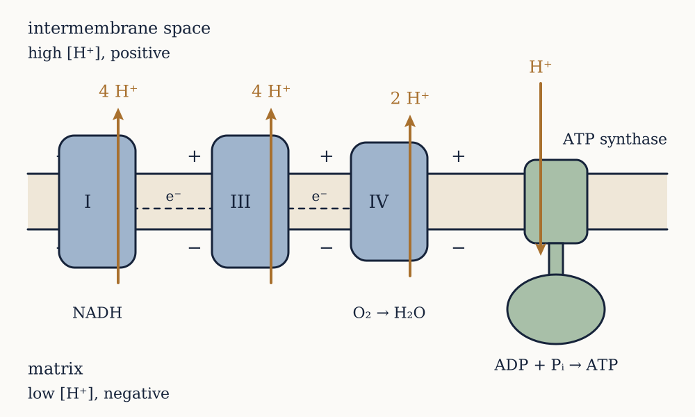
The same principle appears everywhere. Nerve terminals reload neurotransmitter using a proton gradient across the vesicle membrane. Bacteria run flagellar motors on protons rather than on ATP. And mitochondria, as a later lesson shows, turn the entire logic around: instead of spending ATP to build a gradient, they spend a gradient to build ATP.

Every mechanism in this lesson ended at the same place. Channels are free, but the gradients they discharge were not; carriers are free, but they only equalise; pumps cost ATP outright; and cotransporters spend gradients that a pump paid for. The whole traffic across a cell membrane is financed by one molecule, and the next lesson asks what that molecule actually is, why its hydrolysis is worth 50 kJ/mol rather than the 30.5 kJ/mol in the tables, and how a cell keeps the account from ever going empty.

The price of everything

Every process in the last two lessons ended with a bill in ATP, and the bill can only be understood by asking what makes any chemical change happen at all.

This lesson is the thermodynamics the rest of the course spends. It assumes nothing beyond the idea that energy is conserved, and it derives what it needs. Readers who have met free energy elsewhere can skim the first section and start at the one about the phosphate bond, which is where the biology begins and where the standard story is wrong.

What decides whether a reaction goes

Two things change when a reaction happens: the energy stored in bonds, and the number of ways the system and its surroundings can be arranged. The first is the enthalpy change ΔH, negative when heat is released. The second is the entropy change ΔS, positive when the arrangement becomes more disordered. Neither alone predicts direction. Ammonium nitrate dissolves in water spontaneously while absorbing heat, so a reaction can be uphill in enthalpy and still happen, driven by entropy.

The combination that does predict direction, at constant temperature and pressure, is the Gibbs free energy

ΔG=ΔH-TΔS

A process runs forward when ΔG is negative, runs backward when it is positive, and is at equilibrium when it is zero. This is the second law in the form a chemist can use: ΔG negative for the system is exactly the condition for the total entropy of system plus surroundings to increase.

The essential further point is that ΔG depends on concentrations, not only on the identity of the reactants. For a reaction with reaction quotient Q, the ratio of product to reactant activities as they actually are,

ΔG=ΔG+RTlnQ

where ΔG is the value under a defined standard state: one molar for everything, pH 7, 25 degrees Celsius. Nothing in a cell is at one molar and pH 7 is only approximately right, so tabulated ΔG values are a reference point rather than a description. Mistaking one for the other is the commonest error in cellular energetics, and the next section is built on the difference.

One thing ΔG never tells you is speed. Glucose in air has a ΔG of oxidation near -2870 kJ/mol and sits in a sugar bowl indefinitely, because the path to the products runs over an activation barrier. Enzymes lower that barrier and change nothing about the equilibrium. Thermodynamics says what is allowed and catalysis says what happens this afternoon, and a cell needs both.

The bond that is not special

ATP is adenosine with three phosphates in a chain. Hydrolysing the terminal one gives ADP and inorganic phosphate:

ATP+H2OADP+Pi

with ΔG=-30.5 kJ/mol.

Textbooks have called this a "high energy phosphate bond" for eighty years and drawn it with a squiggle, and the phrase is actively misleading. Breaking a bond always costs energy. There is no bond anywhere that releases energy by breaking. What releases energy here is the whole reaction, products against reactants, and three separate features of the products are responsible.

First, electrostatics. At pH 7 the triphosphate chain carries about four negative charges within a few tenths of a nanometre of each other, and they repel. Splitting the chain relieves that strain, and it is the reactant being unstable rather than the products being unusually stable.

Second, resonance. Free inorganic phosphate spreads its negative charge over four equivalent oxygens, an arrangement unavailable to the same phosphate while it is tied into a chain. That delocalisation is worth real stabilisation energy.

Third, solvation. Two separate ions are hydrated better than one larger one, so water is happier with the products.

None of this makes ATP special among phosphate compounds. Phosphoenolpyruvate hydrolyses at -61.9 kJ/mol, twice ATP's figure, and creatine phosphate at -43.1 kJ/mol. ATP sits deliberately in the middle of the range, which is what allows it to be a currency: compounds above it can phosphorylate ADP to make ATP, and ATP can phosphorylate compounds below it. A currency has to be worth less than what you sell and more than what you buy.

Mass action does most of the work

Now put the cell's real concentrations into the equation, which almost no textbook diagram does. A typical cytosol holds roughly 3 mM ATP, 0.3 mM ADP and 5 mM inorganic phosphate. The reaction quotient, in molar units, is

Q=[ADP][Pi][ATP]=(3×10⁻⁴)(5×10⁻³)3×10-3=5×10⁻⁴

and at 310 K, where RT=2.577 kJ/mol,

ΔG=-30.5+2.577ln(5×10⁻⁴)=-30.5-19.6=-50.1 kJ/mol

The real figure is about -50 kJ/mol, sixty per cent larger in magnitude than the tabulated one, and the whole of that extra came from concentrations. This is the number the previous lesson used to show that one ATP is enough to run one cycle of the sodium potassium pump, and with the tabulated 30.5 it would not have been.

The distance from equilibrium can be quantified. The equilibrium constant for ATP hydrolysis is K=e30.5/2.577=1.4×105, while the cell's actual mass action ratio is 5×10⁻⁴. The cell holds ATP about 2.8×10⁸ times away from where the reaction wants to be. At true equilibrium essentially every ATP molecule in you would be ADP.

That is the real nature of ATP. It is not a battery with energy stored in a bond. It is a molecule kept enormously far from its own equilibrium by continuous resupply, and its useful free energy is the size of that displacement. Stop the resupply and the value of the currency collapses within seconds, which is precisely what happens when a tissue loses its blood supply.

Example. A student proposes that a cell could get more energy per ATP by evolving a version whose hydrolysis has a ΔG of -80 kJ/mol instead of -30.5. What would go wrong?

Two things, and they are both about ATP's job rather than its energy content. First, the compound has to be makeable. ATP is regenerated by transferring phosphate from donors such as phosphoenolpyruvate at -61.9 kJ/mol, and no ordinary metabolite could phosphorylate a molecule whose own hydrolysis is worth -80, so the cell would have to find a much stronger donor and would face the same problem one step further back. Second, the compound has to be stable enough to keep. A larger driving force for hydrolysis means a greater tendency to react with water, and ATP's non-enzymatic half-life in neutral solution is already only hours to days. A molecule that hydrolysed spontaneously in minutes would be useless as a store, because a cell recycles its entire ATP pool in about a minute but individual molecules must survive the trip. The general principle is that a currency is defined by sitting between what earns it and what spends it, and moving it to one extreme of the range breaks half of its transactions.

Now you. Sprinting muscle exhausts its ATP in roughly two seconds, yet a sprinter runs for ten. Muscle also holds creatine phosphate at about five times the concentration of ATP, and its hydrolysis is worth -43.1 kJ/mol. What is creatine phosphate for, and why is it not simply used as the currency instead?

Answer

It is a buffer, not a currency. Because its phosphate transfer potential is higher than ATP's, the enzyme creatine kinase can regenerate ATP from ADP directly and almost instantly, with no metabolism involved at all, so the ATP concentration in a working muscle barely falls even while the demand is enormous. Five times the pool at a comparable energy per molecule extends the supply from about two seconds to about ten, which is the duration of a sprint, and it explains why creatine supplementation has measurable effects on short maximal efforts and none on endurance. It is not used as the currency because a currency must be recognised by thousands of different enzymes: ATP is a substrate for kinases, polymerases, motors, pumps and synthetases, and its adenine and ribose give it a large distinctive surface for those enzymes to bind. Creatine phosphate is small and featureless, good for one fast transfer to one partner. The division of labour is between a universally recognised token and a local reserve that can be converted into it.

A currency, not a store

The arithmetic of turnover makes the point better than any argument. Intracellular water in an adult is roughly 28 litres, and at 3 mM the total ATP in a human body is 3×10-3×28=0.084 mol, which at 507 g/mol is about 43 g. Call it fifty grams.

Now the daily consumption. A 2000 kcal diet is 8368 kJ. If something like half of that passes through ATP, at 50 kJ per mole, the body makes and spends 8368×0.5/50=84 mol per day, which is 42 kg of ATP. The efficiency assumption is soft and the honest range is roughly 30 to 60 kg, but every version of the calculation lands at the same qualitative result: an adult turns over something close to their own body mass in ATP each day while never holding more than about fifty grams of it.

Divide the two figures and each ATP molecule is recycled about a thousand times a day, once every minute and a half. There is no reservoir. The system is a pipeline running at full rate, which is why cardiac arrest damages the brain in minutes rather than hours, and why every remaining lesson on metabolism is about the rate at which ATP can be regenerated rather than about how much energy a fuel contains.

Coupling, and why it is mechanical rather than clerical

The most common misconception in bioenergetics is that an unfavourable reaction can be driven by a favourable one simply because the sum of the two free energies is negative. It cannot. Two reactions in the same beaker with no connection between them each go their own way, and the exergonic one merely warms the solution.

Coupling requires a shared intermediate: the two reactions must be steps of a single mechanism, so that the unfavourable one physically cannot happen except as part of the favourable one.

Hexokinase, the first enzyme of glycolysis, is the clean example. Attaching a phosphate to glucose,

glucose+Piglucose 6-phosphate+H2O

has ΔG=+13.8 kJ/mol, so at standard concentrations the equilibrium ratio of product to reactant is e-13.8/2.577=4.7×10-3, and less than half a per cent of the glucose would be phosphorylated. The cell instead runs

glucose+ATPglucose 6-phosphate+ADP

with ΔG=13.8-30.5=-16.7 kJ/mol and an equilibrium ratio of e16.7/2.577=6.5×102. The shift is a factor of 1.4×10⁵, which is exactly the equilibrium constant of ATP hydrolysis, as it has to be.

The crucial detail is that free inorganic phosphate never appears. Hexokinase binds glucose and ATP side by side and transfers the terminal phosphate directly from one to the other in a single step. There is no moment at which ATP has been hydrolysed and the glucose has not yet been phosphorylated, which is what makes the coupling mechanical rather than a piece of accounting. The enzyme also closes around the glucose as it binds, excluding water from the active site, because water is a small and abundant alternative acceptor and an enzyme that let it in would be an ATPase rather than a kinase.

Example. Glutamine synthetase attaches ammonia to glutamate, a reaction with ΔG=+14 kJ/mol, and it uses one ATP. The enzyme does not transfer the phosphate to ammonia. Where must it go, and why does the answer follow from the requirement for a shared intermediate?

It must go onto the glutamate, and it does: the enzyme first makes gamma-glutamyl phosphate, then ammonia attacks that. The reasoning is that the shared intermediate has to lie on the path to the product, so the phosphate must activate the partner that is going to be attacked, not the attacking group. Phosphorylating ammonia would create a dead end, since the cell would then have to get the phosphate off again and nothing would have been gained. The general pattern is worth carrying forward, because it recurs throughout metabolism: ATP is used to convert a poor leaving group, in this case the hydroxyl of a carboxylic acid, into a good one, in this case phosphate. Aminoacyl-tRNA synthetases do the same thing to attach an amino acid to its tRNA, and fatty acid activation does it with coenzyme A. When you see ATP consumed in a biosynthesis, the useful question is not "where did the energy go" but "which group was made into a better leaving group".

Now you. Some biosynthetic reactions consume ATP by splitting it into AMP and pyrophosphate rather than ADP and phosphate, and the cell then immediately hydrolyses the pyrophosphate to two phosphates with a separate enzyme. Why bother with the second step?

Answer

Because it makes the first step effectively irreversible. Splitting ATP to AMP and pyrophosphate is worth about the same as splitting it to ADP and phosphate, roughly -30 kJ/mol under standard conditions, so on its own it buys nothing extra. Destroying the pyrophosphate afterwards, worth a further -19 kJ/mol, removes one of the products of the first reaction, and by mass action a reaction whose product is continuously removed cannot run backwards. The cell is spending a second phosphoanhydride bond purely to buy directionality. This matters most where a mistake would be expensive: DNA and RNA polymerases, aminoacyl-tRNA synthetases and fatty acid activation all use the AMP route, and all of them are committing steps in the assembly of something large. Pyrophosphatase is one of the busiest enzymes in the cell for this reason, and its job is not metabolism but the enforcement of one-way traffic.

The other currencies

ATP is the main one but not the only one, and the others are worth naming because later lessons use them without ceremony.

GTP is chemically almost identical to ATP and is used where a distinct signal is wanted: protein synthesis, the G proteins of signalling, and one step of the citric acid cycle. Interconversion with ATP is fast, so it is not a separate energy pool so much as a separate label.

NADH and NADPH carry electrons rather than phosphate, delivering a hydride ion to whatever needs reducing. They differ by a single phosphate on the ribose, a group that carries no energy and exists purely so that enzymes can tell them apart, and the cell keeps them in opposite states: NADH is held at roughly one part in a thousand relative to NAD⁺, an oxidising pool suited to stripping electrons off fuel, while NADPH is held at around a hundred to one over NADP⁺, a reducing pool suited to building things. One extra phosphate group lets a cell run oxidation and reduction at full tilt in the same compartment at the same time.

Acetyl-CoA carries an activated two-carbon unit as a thioester, whose hydrolysis is worth about -31 kJ/mol, comparable to ATP. And the proton gradient across a membrane, which the previous lesson treated as an expense, turns out in mitochondria to be the largest energy currency of all.

What a cell spends it on

Rough figures make the priorities visible. Making one peptide bond costs four high-energy phosphate bonds, so an average protein of 300 residues costs about 1200 ATP, roughly 60 kJ per mole of protein, before the amino acids themselves are made. A fast-growing bacterium spends the majority of its total energy budget on protein synthesis, with most estimates falling between sixty and seventy-five per cent. Maintaining ion gradients, as the previous lesson noted, takes twenty to thirty per cent of the energy of a resting animal cell and more in neurons. Everything else, including DNA replication, is small by comparison.

So a cell is an object that spends nearly all of its income on making proteins and on keeping sodium outside. Both are continuous, neither can be paused, and the supply has to run at the rate of the demand.

Example. The human brain is about 1.4 kg, two per cent of a 70 kg body, and consumes roughly twenty per cent of resting energy. Work out its energy use per gram against the body average, and say what the difference is spent on.

Twenty per cent of 8368 kJ is 1674 kJ a day, or about 400 kilocalories, which per gram of tissue is 1674/1400=1.20 kJ. The whole-body average is 8368/70000=0.120 kJ per gram. The brain runs at ten times the average rate of the tissue around it. At 50 kJ per mole of ATP that is roughly 17 moles of ATP a day for the organ alone. What it is spent on is mostly the previous lesson's subject rather than this one's: estimates attribute the majority of the brain's energy budget to restoring the sodium and potassium gradients discharged by synaptic and action potential activity, with a further large share to packaging and recycling neurotransmitter. Thinking is expensive because it is electrical, and electrical signalling in a cell means moving ions across a membrane that a pump then has to move back.

Now you. Given that figure, why does concentrating hard on a difficult problem not measurably increase how much you eat?

Answer

Because almost all of the brain's consumption is a standing cost rather than a task cost. The gradients have to be maintained whether or not anything interesting is happening, and the resting firing rates of neurons are already high, so the marginal energy of a demanding task is small against that baseline. Imaging studies that measure regional blood flow and metabolism find task-related increases of a few per cent in the activated regions, which is a large signal locally and a negligible one against total body expenditure of 2000 kilocalories: a few per cent of 400 kilocalories is under 20, which is a biscuit and lies well inside the day-to-day noise in what anyone eats. The wider lesson is one worth carrying into any energy argument about cells. A large total is not the same as a large variable component, and metabolic regulation acts on the part that varies.

Which raises the question this lesson has deferred throughout. If ATP is held 2.8×10⁸ times from equilibrium by continuous resupply, what does the resupplying? The next lesson takes the oldest and most widespread answer, a ten-step pathway that every domain of life still runs, and follows the accounting to an uncomfortable conclusion: it captures almost none of the energy available in the fuel it consumes.

Glycolysis and fermentation

The oldest way a cell makes ATP takes a six-carbon sugar apart into two three-carbon pieces and nets two ATP from the process, which turns out to be a strikingly poor return.

The previous lesson established that ATP is a currency held far from equilibrium by continuous resupply, worth about 50 kJ/mol inside a cell. This lesson is the first supply route: glycolysis, ten enzymatic steps in the cytosol, present in bacteria, archaea and eukaryotes alike, and requiring no oxygen, no membrane and no organelle. Its universality is itself an argument that it is very old.

The shape of the pathway

Glycolysis converts one glucose into two pyruvate, and it does so in two halves that behave quite differently.

The first five steps are a preparatory phase, and they cost money. Hexokinase phosphorylates glucose using one ATP. An isomerase converts glucose 6-phosphate to fructose 6-phosphate. Phosphofructokinase spends a second ATP to make fructose 1,6-bisphosphate. Aldolase then cuts that six-carbon molecule in half, and a final isomerase makes the two halves identical, so the cell now holds two molecules of glyceraldehyde 3-phosphate and is two ATP poorer than when it started.

The last five steps are the payoff phase, and everything after the split happens twice. Glyceraldehyde 3-phosphate dehydrogenase oxidises the aldehyde and captures the energy released as a phosphate bond, producing 1,3-bisphosphoglycerate and reducing NAD⁺ to NADH. Phosphoglycerate kinase hands that phosphate to ADP, making the first ATP. Two rearrangements follow, a mutase and then enolase, which produce phosphoenolpyruvate. Pyruvate kinase hands its phosphate to ADP as well, making the second ATP, and leaves pyruvate.

Two ATP made per three-carbon fragment, twice over, is four; minus the two invested, the net is two ATP and two NADH per glucose. That is the entire yield.

Why spend ATP to get ATP

Investing two ATP before earning any looks perverse and is doing three jobs at once.

It traps the substrate. Glucose enters a cell through a transporter that works in either direction, and glucose could leave the same way. Glucose 6-phosphate carries a charge and cannot cross a membrane or pass back through GLUT, so phosphorylation is a one-way door. It also keeps the internal free glucose concentration low, which keeps the transporter running inward.

It commits the molecule. The phosphofructokinase step is the pathway's true commitment point, because everything before it can be diverted to other uses and nothing after it can, and that is exactly where the regulation sits.

And it destabilises the sugar. Putting phosphate groups at both ends of fructose 1,6-bisphosphate sets up a molecule that aldolase can cleave cleanly into two phosphorylated three-carbon pieces. Cutting an unphosphorylated sugar in half would produce fragments with nothing to hold on to and nothing to activate them.

Where the ATP actually comes from

Glycolysis makes ATP by substrate-level phosphorylation: a phosphate group is handed directly from a metabolite to ADP by an enzyme, with no membrane and no gradient involved. For that to work the donor must have a phosphate transfer potential above ATP's, and glycolysis manufactures two such donors.

The first is built in the glyceraldehyde 3-phosphate dehydrogenase step, and it is the cleverest chemistry in the pathway. Oxidising an aldehyde to a carboxylic acid releases roughly 43 kJ/mol, and in a bomb calorimeter that would all become heat. The enzyme instead attacks the aldehyde with a cysteine thiol to form a thioester, oxidises it there with NAD⁺, and then lets inorganic phosphate displace the thioester. The product, 1,3-bisphosphoglycerate, is an acyl phosphate whose hydrolysis is worth -49.3 kJ/mol, comfortably above ATP's -30.5. Phosphoglycerate kinase then transfers that phosphate to ADP with ΔG=-49.3+30.5=-18.8 kJ/mol, favourable and effectively irreversible under cellular conditions. The energy of an oxidation has been captured as a phosphate bond rather than lost as heat, and note that the phosphate came from free inorganic phosphate in solution, not from any ATP.

The second donor is phosphoenolpyruvate, whose hydrolysis is worth -61.9 kJ/mol, twice ATP's. The reason is not the phosphate but what happens after it leaves: the enol that remains immediately tautomerises to the far more stable keto form of pyruvate, and that rearrangement, which has nothing to do with phosphorus, supplies most of the driving force. Enolase's job in the previous step is simply to create a molecule that is trapped in its enol form by the phosphate group, storing the tautomerisation until a kinase can spend it.

Example. Glucose fully burned to carbon dioxide and water releases 2870 kJ/mol. Fermenting it to two lactate releases 196 kJ/mol. Glycolysis captures two ATP. Assess how good the pathway is, using both the tabulated and the cellular value of ATP.

Two ATP at the standard 30.5 kJ/mol is 61 kJ. Against the 196 kJ that fermentation actually releases, that is 61/196=31 per cent captured, which is respectable and comparable to a petrol engine. Against the 2870 kJ that was available in the glucose, it is 61/2870=2.1 per cent. Using the cellular value of about 50 kJ per ATP the second figure rises to 100/2870=3.5 per cent, and neither version changes the verdict. The two ratios say different things and both are worth holding. Glycolysis is efficient at what it attempts and attempts almost nothing: it takes glucose only as far as pyruvate or lactate, molecules that are still highly reduced and still burnable, so around ninety-six per cent of the chemical energy walks out of the pathway untouched. The reason is visible in the chemistry. Only one oxidation happens in the whole of glycolysis, the GAPDH step, and a carbon atom can be oxidised much further than that. Getting the rest requires stripping every hydrogen off the carbon skeleton, and that needs a terminal electron acceptor the pathway does not have.

Now you. A yeast fermenting sugar to ethanol and a muscle fermenting it to lactate both net exactly two ATP per glucose, despite making completely different products. What does that tell you about where the ATP is made, and what are the two branches actually for?

Answer

It tells you that all the ATP is made upstream of the branch point, which is pyruvate. Everything from glucose to pyruvate is identical in both organisms, and that is where both substrate-level phosphorylation steps sit, so whatever happens to pyruvate afterwards cannot change the yield. The branches therefore exist for some other reason, and the reason is NAD⁺. Both routes take the NADH that GAPDH produced and reoxidise it: lactate dehydrogenase does it in one step by reducing pyruvate, while yeast first removes carbon dioxide to make acetaldehyde and then reduces that to ethanol. Neither branch yields any energy at all, and both in fact throw away the reducing power that NADH represents. That is the correct way to think about fermentation. It is not an energy-producing process, it is a disposal process that exists so the energy-producing steps upstream can keep running, and the ethanol or the lactate is the bill for it.

The bottleneck nobody sees at first

The NAD⁺ requirement is the constraint that shapes the whole of anaerobic metabolism, and its size is easy to underestimate until it is counted.

A cell holds NAD⁺ and NADH together at roughly 0.5 to 1 mM. Glycolysis consumes two NAD⁺ per glucose. A muscle working hard runs glycolysis at something like 1 mM of glucose per second, so it consumes NAD⁺ at 2 mM per second. Against a pool of 0.5 mM, that is complete exhaustion in a quarter of a second.

NAD⁺ is therefore not a reagent but a catalyst that must be turned over hundreds of times a second, and glycolysis stops dead the moment it is all in the reduced form. This is why fermentation exists, and it explains a fact that otherwise looks like waste: the cell discards a molecule, lactate or ethanol, that still contains almost all the energy it started with, purely to get its NAD⁺ back.

Lactate is not a waste product in the ordinary sense. It leaves the muscle, travels in the blood, and is either oxidised by the heart and by other muscle fibres, which take it up and convert it back to pyruvate, or reconverted to glucose by the liver in the Cori cycle at a cost of six ATP per glucose. It is a way of exporting a metabolic problem to a tissue with more oxygen. It is also worth correcting the folklore: lactate does not cause the muscle soreness felt a day or two after exercise, which is inflammation from microscopic muscle damage, and blood lactate is back to baseline within an hour. The burning felt during hard exercise tracks acidification and other metabolites rather than lactate itself, and in fact lactate production consumes a proton rather than producing one.

Example. Yeast fermenting grape juice stops on its own at around 14 to 16 per cent ethanol, which is why fortified wine has to be fortified. Nothing has run out. What has gone wrong?

The yeast has poisoned itself with its own product, and the mechanism is the subject of the second lesson of this course. Ethanol is a small molecule that partitions readily into a lipid bilayer, and at high concentration it disorders the membrane, increases its permeability, and destroys the ion and proton gradients the cell maintains across it. The membrane stops being a barrier, so the cell can no longer hold a composition different from its surroundings, and it dies. This has two consequences worth drawing out. First, a fermentation product is not just a discarded electron sink, it accumulates in the medium and its toxicity sets a hard ceiling on the process, which is exactly the problem industrial ethanol production spends money on. Second, the ceiling varies with strain: wine yeasts tolerate more ethanol than bakers' yeast does, and the difference is largely in membrane lipid composition, including how much ergosterol they can make. A cell's tolerance to a solvent is a property of its membrane, not of its metabolism.

Now you. Cancer cells often ferment glucose to lactate even when oxygen is plentiful, a pattern Otto Warburg described in the 1920s. Fermentation yields fifteen times less ATP per glucose than full oxidation does. Suggest what could make it worth doing anyway.

Answer

Two answers are well supported and a third is speculative, and it is worth being clear which is which. The first is rate rather than yield: glycolysis has few steps, needs no membrane, and can be scaled up simply by making more enzyme, so it can deliver ATP per unit time far faster than oxidative phosphorylation, which is limited by mitochondrial membrane area. A cell competing to divide cares about ATP per second, not ATP per glucose, and glucose is abundant in a tumour. The second is that a dividing cell needs carbon skeletons and reducing power at least as much as it needs ATP: glycolytic intermediates are the precursors of ribose, serine, glycerol and fatty acids, and running high flux through the pathway while diverting intermediates out of it supplies the building blocks for a new cell. Fermenting is partly a way of keeping the flux high without oxidising the carbon away. The third and less settled suggestion is that the exported lactate acidifies the surrounding tissue in ways that favour invasion and suppress immune cells. Warburg's own conclusion, that the mitochondria of cancer cells are damaged and that this causes cancer, was wrong: most tumour cells have functional mitochondria and use them.

Why a bad pathway is universal

Glycolysis captures about three per cent of the energy in its fuel and every organism on Earth still runs it. Several things explain that.

It works without oxygen, and for most of the history of life there was no oxygen to work with. Free atmospheric oxygen appeared only around 2.4 billion years ago, and a pathway that predates it cannot depend on it.

It is fast and simple. Ten soluble enzymes in the cytosol, no membrane, no compartment, no cofactor more exotic than NAD⁺ and ATP. A cell can raise glycolytic flux enormously just by expressing more enzyme, and a muscle fibre in a sprint does exactly that.

It is a supply depot as much as an energy pathway. Glucose 6-phosphate feeds the pentose phosphate pathway, which makes ribose for nucleotides and NADPH for biosynthesis. Dihydroxyacetone phosphate becomes the glycerol backbone of lipids. 3-phosphoglycerate becomes serine, glycine and cysteine. Pyruvate becomes alanine. A cell that shut down glycolysis would lose the raw material for its own construction, not merely some ATP.

And it is the front end of the aerobic route as well. Everything the next lesson describes begins with the pyruvate that glycolysis delivers, so the pathway was never replaced, only extended.

Regulation, and where the control point sits

A pathway with a committed step will be regulated at that step, and phosphofructokinase is the classic case in all of biochemistry.

It is inhibited by ATP, which is the direct feedback: a cell with plenty of ATP does not need to burn sugar. It is activated by AMP, which is a much better signal of energy shortage than ADP because the enzyme adenylate kinase converts two ADP into one ATP and one AMP, so a small percentage fall in ATP produces a large percentage rise in AMP. Reading a small fractional change on a small pool is a general trick for sensitive control. It is also inhibited by citrate, an intermediate of the citric acid cycle, which reports that the downstream pathway is already saturated, and in the liver it is powerfully activated by fructose 2,6-bisphosphate, a molecule that exists for no purpose except signalling and whose concentration is set by hormones.

Example. A muscle holds 5 mM ATP, 0.5 mM ADP and 0.05 mM AMP, and adenylate kinase keeps [ATP][AMP]/[ADP]2 near 1. Suppose demand causes ATP to fall by ten per cent, with the lost ATP appearing as ADP. What happens to AMP, and why does the enzyme read AMP rather than ATP?

ATP goes from 5 to 4.5 mM, a fall of ten per cent, and ADP goes from 0.5 to 1.0 mM, a doubling. Adenylate kinase then sets [AMP]=[ADP]2/[ATP]=1.02/4.5=0.22 mM, against 0.05 mM before. AMP has risen 4.4-fold from a ten per cent fall in ATP. The reason is arithmetic rather than biological: ATP is the large pool and AMP the small one, so the same absolute movement of adenylate is a small fractional change in the first and a large one in the second, and the squared term in the equilibrium amplifies it further. An enzyme trying to detect energy shortage by measuring ATP would be looking for a ten per cent change against a noisy background, while one measuring AMP sees a fourfold signal. This is a general design rule for any sensor: read the species whose fractional change is largest, which is almost always the least abundant member of a conserved pool.

Now you. Metformin, the most widely prescribed drug for type 2 diabetes, mildly inhibits mitochondrial complex I. Given the example above and what a cell does with an AMP signal, sketch the chain from that inhibition to a metabolic effect.

Answer

Partially inhibiting complex I lowers the rate of ATP regeneration, so ATP falls slightly and, by the amplification just derived, AMP rises substantially. The cell has a dedicated reader for that signal, AMP-activated protein kinase, which is switched on by AMP binding and which then acts as a general low-energy alarm: it activates catabolic pathways such as glucose uptake and fatty acid oxidation and inhibits expensive biosynthetic ones such as fatty acid and cholesterol synthesis, and in the liver it suppresses gluconeogenesis, which is the main way metformin lowers blood glucose. The chain is worth noticing for two reasons. The drug never touches glucose metabolism directly; it acts on the respiratory chain and the effect arrives through an energy signal, in the same indirect way digoxin acted through a shared gradient in an earlier lesson. And the honest caveat is that the AMP-activated kinase route is well supported but is probably not the whole story for metformin, since effects have been reported in cells lacking that kinase, so this should be read as the main proposed mechanism rather than a settled one.

Hexokinase is inhibited by its own product, and pyruvate kinase is regulated too, but neither controls the flux the way phosphofructokinase does. The general lesson carries beyond this pathway: control is exerted where commitment happens, and the regulators are chosen so that the enzyme hears both the local state of the pathway and the global state of the cell.

The unavoidable conclusion of this lesson is the accounting. Pyruvate still holds roughly ninety-six per cent of the energy that was in the glucose, sitting in carbon and hydrogen that have not been oxidised. Getting at it means removing every one of those hydrogens and finding somewhere to put the electrons, and doing that with a soluble enzyme handing phosphate to ADP is not possible: no substrate-level step can capture 200 kJ in one bite. The next lesson takes the electrons instead of the phosphate, spends them on a membrane, and turns the resulting gradient back into ATP, which is a mechanism so unlike anything in this lesson that its author was disbelieved for a decade.

Breathing with a gradient

Pyruvate still holds almost all the energy that was in the glucose, and extracting it requires a mechanism with no resemblance to anything in the previous lesson.

The problem is quantitative before it is chemical. The previous lesson found that glycolysis captures around three per cent of what glucose contains, leaving the rest in carbon and hydrogen that have not been oxidised. Recovering it means two things: stripping every hydrogen off the carbon skeleton, and finding somewhere to put the electrons those hydrogens carry. Both are solved in the mitochondrion, an organelle with two membranes, the inner one folded into cristae that give it roughly five times the area of the outer, and a liver cell holds one to two thousand of them.

Getting the carbon ready

Pyruvate crosses into the mitochondrial matrix and meets the pyruvate dehydrogenase complex, one of the largest enzymes known, a machine of dozens of subunits and several million daltons that carries out three chemical steps without releasing the intermediate. It removes one carbon as carbon dioxide, oxidises what is left, reduces NAD⁺ to NADH, and attaches the remaining two-carbon acetyl group to coenzyme A.

The reaction is effectively irreversible, and that single fact has a consequence people meet without knowing why. Fatty acids are broken down to acetyl-CoA, and acetyl-CoA cannot be turned back into pyruvate, so animals cannot convert fat into glucose. A starving human makes glucose from amino acids and from the glycerol backbone of fats, never from the fatty acids themselves, which is why prolonged fasting costs muscle.

A cycle that makes almost no ATP

Acetyl-CoA enters the citric acid cycle, described by Hans Krebs in 1937 in a paper that Nature rejected. Acetyl-CoA condenses with the four-carbon oxaloacetate to give the six-carbon citrate, and the cycle then removes two carbons as carbon dioxide and performs four oxidations, regenerating oxaloacetate ready for the next acetyl group.

Per turn, the output is three NADH, one FADH₂, one GTP and two carbon dioxide. Each glucose delivers two acetyl-CoA, so per glucose the cycle turns twice.

Look at what that yield actually is. One GTP per turn, two per glucose, is the entire direct energy return of the citric acid cycle, and it is made by substrate-level phosphorylation exactly as in glycolysis. Everything else the cycle produces is reduced electron carriers. The cycle is not an ATP-generating pathway. It is a device for taking carbon apart and loading the electrons onto NAD⁺ and FAD, and the reason it is a cycle rather than a line is that the acceptor, oxaloacetate, has to be regenerated: four carbons in as acetyl, two carbons out as carbon dioxide, and the catalyst back where it started.

The cycle is also a supply depot, in the same way glycolysis is. Citrate leaves the mitochondrion to supply carbon for fatty acid synthesis, alpha-ketoglutarate becomes glutamate, oxaloacetate becomes aspartate. Drawing intermediates out would drain the cycle, so cells maintain separate reactions that top oxaloacetate back up, and running the cycle at all requires keeping that balance.

What an NADH is worth

Now the electrons. The tendency of a couple to accept them is measured by its standard reduction potential, and two values decide everything here: NAD⁺ / NADH sits at -0.32 V, and oxygen reduced to water sits at +0.82 V. The gap is 1.14 V, and the free energy released when two electrons fall across it is

ΔG=-nFΔE=-2×96485×1.14=-220 kJ/mol

At about 50 kJ per ATP inside a cell, one NADH is worth roughly four and a half ATP. That is the prize, and it also states the difficulty. No substrate-level phosphorylation can capture 220 kJ in one transfer, because the largest phosphate transfer potential available is phosphoenolpyruvate at 62 kJ/mol. Trying to take the whole drop in one bite would waste most of it as heat.

So the cell takes it in stages. The inner mitochondrial membrane holds four large complexes arranged in order of increasing reduction potential, and electrons pass down the series like water through a flight of locks. Complex I takes them from NADH. Complex II takes them from succinate, which is why FADH₂ made inside the cycle enters lower down and is worth less. Both hand their electrons to ubiquinone, a small lipid-soluble carrier that diffuses within the membrane to complex III, which passes them to cytochrome c, a small water-soluble protein on the outer face of the inner membrane, which delivers them to complex IV. Complex IV puts four electrons and four protons onto one molecule of oxygen and releases two waters.

That final step is why we breathe. Oxygen is not consumed to burn anything directly; it is the terminal electron acceptor, the sink at the bottom of the flight of locks, and the entire chain backs up if it is missing. Cyanide and carbon monoxide kill by binding the iron in complex IV, which is why they act in seconds: nothing has been poisoned except the last step, and the whole chain stops with it.

Mitchell's heresy

Complexes I, III and IV pump protons across the inner membrane as electrons pass through them: roughly four, four and two protons per pair of electrons, ten in total for each NADH, and six for each FADH₂ since it bypasses complex I.

That this is the mechanism took fifteen years to accept. Through the 1950s the field looked for a "high energy intermediate", a chemical compound analogous to 1,3-bisphosphoglycerate that would be made by the respiratory chain and would then phosphorylate ADP. Dozens of laboratories searched for it and none found it.

Peter Mitchell proposed in a 1961 paper in Nature that there is no such compound, and that the intermediate is not chemical but electrical: the chain uses the energy of electron transfer to pump protons out, creating a gradient across the membrane, and ATP synthase then lets protons back in and uses the flow to make ATP. The proposal was widely rejected. It required the membrane to be intact and impermeable, made the coupling stoichiometry non-integral, and came from a scientist who had left his university post and was working in a converted manor house in Cornwall.

Two experiments settled it, and both are worth knowing because neither is a detail.

André Jagendorf and Ernest Uribe, in 1966, took chloroplast thylakoid membranes, soaked them in acid at pH 4 until the inside equilibrated, then jumped the outside to pH 8 and added ADP and phosphate. In complete darkness, with no light, no electron transfer and no substrate being oxidised, the vesicles made ATP. A pH difference alone was sufficient, which is precisely what the chemiosmotic hypothesis predicts and what no chemical intermediate theory can explain.

Efraim Racker and Walther Stoeckenius, in 1974, built the converse. They made artificial lipid vesicles containing two purified proteins with no evolutionary or functional relationship: bacteriorhodopsin, a light-driven proton pump from a salt-loving archaeon, and mitochondrial ATP synthase from cow heart. Shine light on the vesicles and they make ATP. Nothing connects the two proteins except the proton gradient in between, which is a direct demonstration that the gradient is the entire linkage. Mitchell received the Nobel Prize in 1978.

Example. Someone proposes that ATP synthase is driven by direct physical contact with the respiratory complexes. Which single feature of the Racker and Stoeckenius experiment refutes this, and what would you predict about the spacing of the two kinds of protein in a real mitochondrion?

The refutation is that the two proteins in the vesicle come from different domains of life and have never met in any organism, so no evolved interaction surface can exist between them, and yet the system works. Contact cannot be the mechanism. The prediction that follows is that in a real mitochondrion the respiratory complexes and the ATP synthase need not be adjacent, and they are not: electron microscopy of cristae shows ATP synthase concentrated in rows along the highly curved rims of the folds while the respiratory complexes sit in the flatter membrane, sometimes hundreds of nanometres away. That separation would be inexplicable on a direct-contact model and is unremarkable on a chemiosmotic one, because the gradient is a property of the whole compartment rather than of any location in it. It is also why the mechanism has a requirement no soluble pathway has: the membrane must be closed. Puncture the vesicle anywhere and ATP synthesis stops everywhere.

Now you. Mitchell's hypothesis was resisted partly because it predicted a non-integral number of ATP per NADH, whereas every pathway known at the time gave whole numbers. Why does chemiosmosis produce a fraction, and why is that a point in its favour rather than against it?

Answer

Because the two halves of the process are not chemically joined. In substrate-level phosphorylation one molecule hands one phosphate to one ADP, so the ratio is one by construction. In chemiosmosis the chain pumps some number of protons into a shared pool and the synthase withdraws some other number, and the two numbers are set by entirely separate pieces of machinery: how many protons a complex happens to translocate per electron pair, and how many subunits happen to be in the synthase's rotor ring. There is no reason for one to divide the other. That it is a point in favour becomes clear once the rotor rings are counted, because they differ between organisms: eight subunits in mammalian mitochondria, ten in yeast, up to fifteen in some bacteria, which means the protons per ATP differ between species running otherwise identical chemistry. A chemical intermediate model cannot accommodate that at all, while a gradient model predicts exactly this kind of variation, and predicts further that an organism living on a shallow gradient should have a larger ring. Larger rings are indeed found in organisms with weak proton-motive force.

The size of the gradient

The proton-motive force has two parts, because a proton carries both concentration and charge:

Δp=Δψ-2.3RTFΔpH

In a working mitochondrion the membrane potential is about 160 mV, inside negative, and the matrix is roughly 0.75 pH units more alkaline than the intermembrane space, worth about 45 mV at 37 degrees Celsius. Together Δp is around 200 mV, and it is dominated by the electrical term rather than by the pH difference, which is not the intuitive expectation.

Each proton returning through that gradient can release F×0.200=19.3 kJ/mol. Ten protons per NADH therefore capture 193 kJ of the 220 kJ available, which is 88 per cent, an efficiency at the pumping stage that no engine approaches.

The machine that sells the gradient back

ATP synthase is a rotary motor, and saying so is not a metaphor. The membrane-embedded F₀ portion contains a ring of c subunits which turns as protons pass through the interface between the ring and an adjacent subunit; the central stalk, attached to the ring, rotates inside the mushroom-shaped F₁ head that projects into the matrix, and the head contains three catalytic sites.

Paul Boyer proposed in the 1970s that the sites do not make ATP by pushing the reaction energetically, but by binding change: at any moment the three sites are in different conformations, one loosely binding ADP and phosphate, one tightly bound with ATP already formed, and one open. The remarkable claim, since confirmed, is that forming ATP from ADP and phosphate in the tight site is close to energy-neutral, because the enzyme binds ATP so much more tightly than the substrates that it pays the cost of the bond. The energy from the proton gradient is spent almost entirely on releasing the finished ATP, by rotating the stalk and forcing the site open.

John Walker's crystal structure in 1994 showed the three sites in exactly the three predicted states, and Boyer and Walker shared a Nobel Prize in 1997. Hiroyuki Noji and colleagues then made rotation visible in the same year, by fixing an F₁ head to a glass slide, attaching a fluorescently labelled actin filament to the central stalk, adding ATP, and watching the filament turn under a microscope in discrete 120-degree steps.

The stoichiometry follows from the geometry. One full rotation makes three ATP, one for each catalytic site, and requires as many protons as there are c subunits in the ring. Mammalian mitochondria have eight, so 8/3=2.7 protons per ATP, and roughly one more proton per ATP is spent by the transporters that bring ADP and phosphate into the matrix and carry ATP out, giving about 3.7 in total. At 19.3 kJ per proton that is 71 kJ delivered to make a molecule worth 50 kJ, which is enough with margin, as it must be for the reaction to run at a useful rate.

Example. Making one ATP inside a cell costs about 50 kJ/mol. Using Δp=200 mV, work out the minimum number of protons the synthase must use, and then predict the rotor ring size of a bacterium that lives on a proton-motive force of only 120 mV.

Each proton delivers FΔp=96485×0.200=19.3 kJ/mol, so making one ATP needs at least 50/19.3=2.6 protons, and since a rotor ring cannot deliver fractions per site the minimum practical figure is three. The mammalian ring of eight subunits gives 2.7 per ATP, which is just above that floor, consistent with a mitochondrion running at a healthy gradient and no more. Now repeat at 120 mV: each proton is worth 96485×0.120=11.6 kJ/mol, so an ATP needs 50/11.6=4.3 protons, and a ring making three ATP per revolution must therefore have about 3×4.3=13 subunits. That is a real prediction and it holds: alkaliphilic Bacillus species, which live at high external pH and therefore cannot build a large pH component to their gradient, have rotor rings of thirteen. A structural parameter of a motor can be predicted from the electrochemistry of the environment its owner lives in, which is about as direct a confirmation of a mechanism as biology offers.

Now you. Suppose the inner membrane of a mitochondrion becomes leaky, so the gradient collapses, but the cell still contains plenty of ATP made by glycolysis. What does ATP synthase do, and what would you expect a cell to have evolved in response?

Answer

It runs backwards. The enzyme is a reversible rotary motor and nothing about it enforces a direction; with no gradient to spend, the only remaining source of free energy is ATP hydrolysis, so the machine hydrolyses ATP and pumps protons out, driving the rotor the other way. This is not a curiosity: it is what happens in ischaemic tissue, and it means a cell deprived of oxygen destroys its own glycolytic ATP through its mitochondria unless something stops it. What has evolved in response is a small inhibitor protein, IF1, which binds the catalytic head and blocks the hydrolytic direction while leaving the synthetic direction unaffected. It is activated by the fall in matrix pH that accompanies loss of the gradient, so it switches on exactly under the conditions where reversal would occur. The general shape of the argument recurs throughout this course: any machine that couples two processes reversibly will run whichever way the free energy points, so a cell that needs one direction only has to spend a separate component on enforcing it.

What a glucose is actually worth

Now the whole account, per glucose. Substrate-level phosphorylation gives four ATP, two in glycolysis and two as GTP in the cycle. The reduced carriers are ten NADH, counting two from glycolysis, two from pyruvate dehydrogenase and six from two turns of the cycle, plus two FADH₂. The protons pumped are 10×10+2×6=112.

Divide by 3.7 protons per ATP and the chain yields 30 ATP, for a mechanistic total near 34. Measured ratios come out lower, at about 2.5 ATP per NADH and 1.5 per FADH₂, giving 32 in total, or 30 if the cell uses the cheaper of the two shuttles that carry cytosolic NADH into the mitochondrion. The gap between 34 and 32 is real and is mostly proton leak: the inner membrane is not perfectly impermeable, and some of the gradient is discharged without passing through the synthase.

So the honest figure is "about 30", not a whole number, and its imprecision is a property of the mechanism rather than of the measurement. Older textbooks give 38, which came from assuming three ATP per NADH and two per FADH₂ before anyone had measured the rotor ring.

Compare with the previous lesson. Fermentation nets two ATP per glucose and respiration nets about thirty, a factor of fifteen, and at 50 kJ per ATP the aerobic route captures 32×50/2870=56 per cent of the energy in the fuel. That is the payoff for the entire apparatus of membranes, complexes and gradients.

A cross-check is available and it works. An adult at rest consumes about 250 mL of oxygen per minute, which is 0.25×1440/22.4=16 moles per day. Each oxygen molecule accepts four electrons, that is two NADH, worth about five ATP. So the body should make 16×5=80 moles of ATP a day, or 41 kg. The previous lesson estimated 42 kg from an entirely independent route, dietary calories divided by energy per ATP. Two calculations sharing no inputs landing within a kilogram is the sort of agreement that should raise confidence in both.

Example. 2,4-dinitrophenol is a weak acid that is lipid-soluble in both its protonated and its deprotonated form. Predict its effect on a mitochondrion, and on a person.

Being lipid soluble in both forms, it can pick up a proton on the acidic side of the membrane, diffuse across as the neutral molecule, release the proton on the alkaline side, and diffuse back as the anion. It is a proton shuttle, and it short-circuits the gradient. Electron transport continues, because oxygen is still available and the chain is not blocked, but the protons return without passing through ATP synthase, so the energy emerges entirely as heat and ATP synthesis stops. The prediction for the organism is oxygen consumption rising sharply, body temperature rising, weight falling because fuel is being burned to no purpose, and death from hyperthermia if the dose is high. All of that is documented, because DNP was sold as a slimming drug in the United States from 1933, was taken by an estimated hundred thousand people, caused deaths and cataracts, and was banned in 1938. It is still sold illegally and still kills. The pharmacological lesson is that a drug with no therapeutic ratio is not a drug: the mechanism that produces weight loss is the mechanism that produces fatal hyperthermia, and there is no dose at which one happens without the other.

Now you. Newborn humans and hibernating mammals carry brown adipose tissue, packed with mitochondria and expressing a protein called UCP1 in the inner membrane. Infants cannot shiver. What does UCP1 almost certainly do, and what does its existence say about the argument in this lesson?

Answer

It must be a regulated proton channel that lets protons back into the matrix without passing through ATP synthase, and it is: uncoupling protein 1, activated by fatty acids and inhibited by purine nucleotides, doing deliberately what dinitrophenol does indiscriminately. Fuel is oxidised at full rate and the energy appears as heat rather than ATP, which is how an infant or a hibernator warms itself without muscle activity. What its existence says about the argument is more interesting than the physiology. If the coupling between oxidation and ATP synthesis were a chemical intermediate, a protein that made a membrane leaky to protons could not possibly abolish ATP synthesis, and evolution could not have built a thermogenic tissue by adding one channel. That a single protein, doing nothing but conducting protons, converts a mitochondrion from an ATP factory into a heater is chemiosmosis demonstrated in a living animal. Physiology is here confirming a mechanism that was established in vitro, which is the direction of evidence one always hopes for and rarely gets.

The six lessons so far have built a cell that holds itself apart from the world, moves what it needs across its boundary, and pays for all of it. Every step has been carried out by a protein: a channel with carbonyls spaced to fit a potassium ion, a kinase that excludes water from its active site, a rotor with exactly eight subunits. None of that could be arrived at by chance in the lifetime of a cell, and none of it was designed. It was inherited, as a specification, from the cell that divided to make this one. The next lesson asks where that specification is kept, and the answer took four experiments over twenty-five years to establish, because everybody expected it to be the proteins themselves.

What the gene is made of

Every mechanism in the previous six lessons was a specific protein, inherited as a specification from the cell that divided to make this one, and until 1944 almost everyone assumed the specification was itself protein.

That assumption was reasonable, which is worth saying before it is demolished. Chromosomes are made of protein and DNA together. Proteins are built from twenty different amino acids and can be arranged in astronomically many ways, while DNA has only four bases and looked monotonous. Phoebus Levene, who identified the components of nucleic acid, had proposed that DNA was a repeating tetranucleotide, a dull structural polymer with the four bases in fixed rotation. A molecule with no variety cannot carry a message, and for thirty years the field believed DNA had no variety.

A mouse experiment nobody understood

Frederick Griffith was a public health bacteriologist studying pneumococcus during the pandemics of the 1920s, and he was not looking for genes. The bacterium comes in two forms: a smooth strain with a polysaccharide capsule, which is lethal to mice, and a rough strain without one, which is harmless because white blood cells can engulf it.

His 1928 result was a four-way comparison, and its power comes from the controls rather than from the striking case. Live rough bacteria: the mouse lives. Live smooth: the mouse dies. Heat-killed smooth: the mouse lives, so heat has destroyed the organism. Heat-killed smooth mixed with live rough: the mouse dies, and living smooth bacteria can be recovered from its blood.

Something had passed from the dead cells into the living ones and permanently changed them, and it bred true in the descendants. Griffith called it the transforming principle and did not speculate much about what it was. Notice how little the experiment assumes: no chemistry at all, only the observation that a heritable trait can be transferred between cells by a substance from a corpse.

Purifying the principle

Oswald Avery, Colin MacLeod and Maclyn McCarty spent more than a decade at the Rockefeller Institute reproducing transformation in a test tube and then asking which chemical fraction did it. Their 1944 paper is a model of subtractive reasoning.

They prepared extracts of heat-killed smooth cells and purified the active material. Its chemistry looked like DNA: the ultraviolet absorption, the elemental ratio of nitrogen to phosphorus, and behaviour on precipitation with alcohol all matched. Then the removals. Proteases destroyed the protein and transformation still worked. Ribonuclease destroyed the RNA and transformation still worked. Lipid extraction changed nothing. Deoxyribonuclease abolished it completely.

The logical structure is clean: everything else can be destroyed without effect, and only the one thing cannot. The reception was not. The dominant objection was that a trace of protein could have survived the treatment and been the real agent, since a very small amount of a very active substance would be undetectable, and Alfred Mirsky pressed this in print for years. Avery himself was cautious in a way the paper's later reputation obscures, and he was never awarded a Nobel Prize.

The objection was not unreasonable. It is genuinely hard to prove that a purified preparation contains none of a contaminant, and the counter-argument in the end was not chemical purity but the accumulation of independent lines of evidence.

The blender

Alfred Hershey and Martha Chase gave one of those lines in 1952, using a virus that infects bacteria. Bacteriophage T2 consists of nothing but protein and DNA, and its life cycle offered a natural separation: the phage attaches to the outside of the bacterium and something enters, after which the cell produces hundreds of new phage.

The trick is that protein and DNA can be labelled separately by elements each contains and the other does not. Protein contains sulfur, in cysteine and methionine, and no phosphorus. DNA contains phosphorus in its backbone and no sulfur. So they grew one batch of phage with radioactive sulfur-35 and another with radioactive phosphorus-32, let each infect bacteria, then sheared the attached phage coats off the cell surfaces in a kitchen blender and spun the mixture so that the heavy bacteria pelleted and the light phage coats stayed in the supernatant.

Most of the sulfur label ended up in the supernatant, with the discarded coats. Most of the phosphorus label stayed with the cells, and around thirty per cent of it appeared in the next generation of phage. Protein stays outside, DNA goes in.

Example. In the Hershey and Chase experiment roughly twenty per cent of the sulfur label stayed with the bacteria and a substantial fraction of the phosphorus was washed off. Given that the folklore version reports a clean separation, how much weight should the experiment carry?

Less than it is usually given, and it is worth being precise about why. The result is a difference in distribution, not a clean partition: some protein enters or sticks, some DNA fails to. Hershey's own paper is correspondingly hedged, and it concludes that the protein has no function in growth rather than that DNA is the genetic material. Taken alone the experiment is suggestive rather than decisive, and a determined critic could have argued that the twenty per cent of protein entering the cell was the important twenty per cent. What made it persuasive was not its internal cleanliness but its independence: it used a different organism, a different technique and a different logic from Avery's, and it pointed the same way. That is the honest structure of the case for DNA. No single experiment closed it. Three did, together with the fact that the structure found the following year immediately explained something no protein model could. It is also a useful correction to how experiments get remembered, because the blender is memorable and the decade of careful enzymology at the Rockefeller is not.

Now you. Suppose Avery, MacLeod and McCarty's preparation really had contained a trace of protein that was the true transforming agent. Their deoxyribonuclease result would then need another explanation. Can you construct one, and what would you do to test between the two?

Answer

A defender of protein could argue that the hypothetical active protein is bound to DNA and requires that DNA as a carrier or scaffold, so destroying the DNA would inactivate it without the DNA itself being the message. The argument is not absurd, since DNA-binding proteins are real. Two tests distinguish the possibilities. The first is dose response and specificity of the enzyme: deoxyribonuclease abolishes activity while proteases at high concentration do not, and if the carrier account were right one would expect at least partial loss with proteases too, since a protein stripped from its scaffold should also fail. The second and stronger test is to make the material rather than purify it. Transformation with chemically synthesised DNA of known sequence, or with DNA that has passed through a step no protein could survive, removes the contamination argument entirely. That is essentially what the field eventually did, and it is the general escape from any purification argument: stop subtracting and start constructing, because a contaminant that survives a synthesis you performed yourself is not a contaminant anyone can appeal to.

Chargaff's ratios

Erwin Chargaff, at Columbia, applied paper chromatography to hydrolysed DNA from many organisms and published in 1950 two findings, one of which is famous and one of which mattered more at the time.

The famous one is that within any species the amount of adenine equals the amount of thymine and the amount of guanine equals the amount of cytosine. In human thymus DNA the four bases come out at roughly 30.9, 29.4, 19.9 and 19.8 per cent, so A/T=1.05 and G/C=1.005. Within experimental error, one to one.

The finding that mattered more in 1950 is that the ratio of the AT pairs to the GC pairs varies enormously between species. Human DNA is about 40 per cent G plus C, Plasmodium falciparum around 20 per cent, some Streptomyces over 70. That range killed the tetranucleotide hypothesis outright. DNA is not a monotonous repeat; its composition differs from organism to organism, which is the minimum requirement for carrying information.

Example. A bacterium's DNA is 62 per cent G plus C. Work out the percentage of each of the four bases, and say what you can and cannot infer from a high figure.

Chargaff's rules give G=C and A=T, so the 62 per cent divides equally into 31 per cent guanine and 31 per cent cytosine, and the remaining 38 per cent gives 19 per cent adenine and 19 per cent thymine. What you can infer is a physical property: a GC pair makes three hydrogen bonds and an AT pair two, and GC-rich DNA also stacks more favourably, so a GC-rich genome has a higher melting temperature, which is the basis of every calculation anyone does when designing a primer. What you cannot safely infer is that the organism lives somewhere hot. The correlation between whole-genome GC content and growth temperature across prokaryotes is weak, and the strong correlation is with the GC content of the paired stems of ribosomal and transfer RNA, which are structural molecules that must stay folded. Genomic GC content correlates better with other things entirely, including which repair and mutational biases the lineage has. This is a good instance of a real physical mechanism supporting an inference at one level and not at another.

Now you. Two viral genomes are analysed. The first gives A 32, T 32, G 18, C 18 per cent. The second gives A 25, T 33, G 24, C 18. What is the most important difference, and what would you conclude?

Answer

The first obeys Chargaff's rules, with A=T and G=C, and the second does not: adenine and thymine differ by eight percentage points and guanine and cytosine by six. Since the rules follow from every base being physically paired with its complement along a double-stranded molecule, a genome that violates them is not double-stranded. The conclusion is that the second virus has a single-stranded genome, and this is exactly how such genomes were first recognised, in the phage φX174, whose measured composition is close to the second set of numbers. Two further points are worth taking. This is a case where a rule is more useful for its exceptions than for its instances, since the rule holding tells you only what you already assumed while the rule failing identifies something new. And the inference is quantitative rather than qualitative, so it requires knowing the measurement error: base composition determined by chromatography carried an uncertainty of a per cent or so, which is why a discrepancy of six to eight points is convincing and one of one point would not be.

Chargaff had the essential clue and did not see what it meant. The pairing rules are a chemical fact in search of a structural explanation, and supplying that explanation is what the following three years were about.

The structure, and who supplied the data

Rosalind Franklin and Maurice Wilkins at King's College London were taking X-ray diffraction photographs of DNA fibres. Franklin, with her student Raymond Gosling, obtained in May 1952 the photograph known as Photo 51, of the hydrated B form, and it is one of the most information-dense images in science.

Three things can be read off it almost directly. The bold X-shaped cross of reflections is the diffraction signature of a helix, and the angle of its arms gives the pitch relative to the diameter. The spacing of the layer lines gives a repeat of 3.4 nm along the fibre axis. And a strong reflection at 0.34 nm gives the spacing between successive stacked bases, so a turn of the helix contains 3.4/0.34=10 base pairs. The missing fourth layer line indicates two strands offset from each other rather than one, which is where the major and minor grooves come from. Franklin's own analysis had also established that the phosphate backbone lies on the outside, which ruled out the three-chain models with the bases outward that Linus Pauling published and that James Watson and Francis Crick had earlier attempted.

Watson and Crick, at Cambridge, built models rather than taking data, and in early 1953 they saw Photo 51 and an unpublished Medical Research Council progress report containing Franklin's numerical results. This was done without her knowledge or agreement. Their paper in Nature on 25 April 1953 cites her only as having "unpublished" work that "stimulated" them, and she died of ovarian cancer in 1958 at 37, four years before the Nobel Prize went to Watson, Crick and Wilkins. The structure is correct and the model-building was a genuine achievement; the credit was not distributed honestly, and any account that omits this is telling the story wrong.

Why the pairing has to be what it is

The model's central constraint is uniform width, and this is where Chargaff's ratios stop being a coincidence.

The bases come in two sizes. Adenine and guanine are purines, double-ring systems. Thymine and cytosine are pyrimidines, single rings and roughly half the width. If the two backbones are to run at a constant separation, every rung of the ladder must be the same length, and the only way to achieve that with these four components is to pair one purine with one pyrimidine. Two purines would bulge, two pyrimidines would pinch, and either would kink the helix.

That narrows it to four possible pairings, and hydrogen bonding chooses between them. In their correct tautomeric forms, adenine presents a donor and an acceptor in positions that match thymine exactly, making two hydrogen bonds; guanine and cytosine match at three positions. Adenine against cytosine puts two acceptors opposite each other and does not bond. So A pairs with T and G with C, and this is precisely Chargaff's result: the ratios are one to one because the bases are physically paired one to one along the molecule.

The geometry has a further consequence that is easy to pass over and is the reason DNA can carry information at all. Because every rung has the same width, any sequence of pairs makes an equally good helix. The molecule imposes no constraint on the order of its own letters. Compare a protein, whose sequence determines whether it folds at all, and where most random sequences are useless. DNA is a structurally indifferent medium, which is exactly what a message wants to be written on.

Count the capacity. Four possibilities per position is two bits, so the E. coli genome of 4,641,652 base pairs holds 9.3×106 bits, about 1.16 megabytes. A haploid human genome of 3.2 billion base pairs holds 800 megabytes. These figures are upper bounds, since real genomes are highly redundant and much of a human genome is repeated sequence, but they establish the order of magnitude: everything needed to specify a human being would fit on a compact disc.

Example. A student says the two DNA strands carry the same information twice over, so the molecule is fifty per cent redundant and evolution should have removed the duplication. What is wrong with this?

Nothing about the fact, and everything about the conclusion. The two strands are indeed redundant in the information-theoretic sense: given one, the other is fully determined by the pairing rules, so the molecule stores 2 bits per base pair rather than 4. But redundancy is what the structure is for. It gives a copying mechanism, since each strand is a template for the other, which is the subject of the next lesson. It gives a repair mechanism, since damage to one strand can be corrected by reading the intact partner, and a cell suffers thousands of DNA lesions a day that would be uncorrectable in a single-stranded molecule. And it gives chemical stability, because the reactive base edges are turned inward and stacked, protected from water and from oxidation. Single-stranded genomes do exist, in many viruses, and they show the expected properties: much higher mutation rates and much smaller genomes. RNA viruses run mutation rates around 10-4 per base per replication against 10-9 for a cell, and no RNA virus has a genome much beyond thirty thousand bases. The redundancy is not waste. It is what makes a large genome possible.

Now you. Photo 51 shows a strong reflection at a spacing of 0.34 nm and a repeat of 3.4 nm, giving ten base pairs per turn. Suppose an X-ray photograph of a different nucleic acid gave 0.28 nm and 3.1 nm instead. What would you conclude, and what would you want to check next?

Answer

You would conclude that the bases are stacked more closely, at 0.28 nm, and that the helix contains 3.1/0.28=11 base pairs per turn, so it is a different helical form: more tightly wound and shorter per base. This is close to the real A form of DNA, which is what B-DNA becomes when it is dehydrated, and it is also the form double-stranded RNA takes, since the extra hydroxyl on ribose prevents the sugar from adopting the conformation B-DNA requires. The check to run next is humidity, which is exactly what the King's group did: Franklin's key experimental contribution was recognising that DNA fibres exist in two distinct forms and that the sharp, interpretable pictures came from the hydrated B form, so controlling water content was the difference between an uninterpretable smear and Photo 51. The wider point is that a diffraction pattern gives dimensions rather than a structure, and turning dimensions into a structure needs chemistry: bond lengths, ring geometry, what can hydrogen bond to what. Watson and Crick's contribution was that second step, and it needed the first.

The sentence at the end of the paper

Watson and Crick closed their 1953 paper with one of the most quoted sentences in science: "It has not escaped our notice that the specific pairing we have postulated immediately suggests a possible copying mechanism for the genetic material."

The claim is that the structure does not merely accommodate heredity, it explains it. Separate the two strands, and each carries the complete information needed to rebuild its partner, because every base specifies what must sit opposite it. A gene is a sequence, a copy is made by templating, and the same base pairing that holds the molecule together is the mechanism by which it is duplicated.

That is a hypothesis, and a strong one, because it makes a prediction that could fail. If each new double helix is built from one old strand and one new one, then after a single round of copying in labelled medium every molecule should be a hybrid, and after two rounds half should be hybrid and half entirely new, in a specific and measurable ratio. Two other schemes were live at the time and predicted different outcomes. The next lesson is the experiment that decided between them, which is often called the most beautiful in biology, and then the machinery that turns out to carry out the copying, which is stranger than the elegant picture suggests.

Copying it

A structure that suggests a copying mechanism is a hypothesis, and by 1957 there were three competing versions of how DNA might be duplicated.

The previous lesson ended with Watson and Crick's remark that base pairing suggests a way to copy the molecule. Take that seriously and three things could happen when a double helix is duplicated. Semiconservative: the strands separate, each templates a new partner, and every daughter molecule has one old strand and one new. Conservative: the original double helix somehow directs the synthesis of an entirely new one and stays intact itself. Dispersive: the molecule is copied in pieces that are interleaved, so both daughter molecules are patchworks of old and new along their length.

All three are consistent with base pairing. Only an experiment can choose.

The most beautiful experiment in biology

Matthew Meselson and Franklin Stahl, at Caltech in 1958, found a way to weigh DNA precisely enough to tell the three apart.

The tool is caesium chloride density gradient centrifugation. Spin a concentrated caesium chloride solution at very high speed for long enough and the heavy caesium ions redistribute until the solution has a smooth density gradient down the tube. DNA in that tube migrates to the depth where the solution density matches its own and forms a sharp band there, which can be photographed by ultraviolet absorption. The resolution is remarkable: it separates molecules differing in density by well under one per cent.

The label is nitrogen. DNA bases are rich in nitrogen, and the heavy stable isotope nitrogen-15 is not radioactive, so bacteria can simply be grown on it. Meselson and Stahl grew E. coli for many generations on ammonium chloride made with nitrogen-15 until essentially all its DNA was heavy, then abruptly transferred the culture to ordinary nitrogen-14 medium and took samples at intervals.

The predictions differ sharply. After exactly one round of replication, conservative copying gives two populations, one fully heavy and one fully light, so two bands. Semiconservative gives every molecule one heavy strand and one light one, so a single band exactly halfway between. Dispersive also gives a single band halfway between, so one generation cannot distinguish the last two.

The observation after one generation was a single intermediate band, at the density expected for a hybrid. Conservative replication was dead.

The second generation separates the survivors. Semiconservative copying predicts that each hybrid molecule gives one hybrid and one fully light molecule, so half hybrid and half light, in two distinct bands. Dispersive copying predicts a single band that has moved to three-quarters light, since every molecule is still a uniform patchwork. What appeared was two bands in equal amounts, one at hybrid density and one at light. Dispersive was dead too.

Meselson and Stahl added a further check that is often left out and which closes the argument properly. They heated the hybrid DNA to separate the strands, and the two single strands banded at two different densities, one fully heavy and one fully light. Each strand was uniform along its length, which no dispersive scheme allows.

Semiconservative replication was established with a design that answers the question directly, needs no assumptions about mechanism, and can be understood from the photograph of the tubes alone.

Why the machinery cannot be as tidy as the picture

The clean image is two strands unzipping while polymerases run along behind them. The actual chemistry forbids it, for a reason worth deriving rather than memorising.

DNA polymerase adds a nucleotide by having the free 3' hydroxyl of the growing chain attack the innermost phosphate of an incoming nucleoside triphosphate, displacing pyrophosphate. The energy for the new bond comes from the incoming nucleotide, not from the chain. Synthesis therefore runs only in the 5' to 3' direction, because that is the end that carries a free hydroxyl to attack with.

Could a polymerase have evolved to run the other way, carrying the triphosphate at the growing end of the chain and attacking incoming nucleotides with it? Chemically, perhaps. But then any removal of a wrongly incorporated base would take the triphosphate away with it and leave a chain that cannot be extended, and the whole proofreading system described below becomes impossible. One-directional synthesis is the price of being able to correct mistakes, and every DNA polymerase in every organism pays it.

The consequence is that the two strands of a replication fork cannot be copied in the same way, because they are antiparallel. As the fork opens, one template runs 3' to 5' into the fork, and its new strand can be made continuously in the direction the fork is moving. This is the leading strand. The other template runs the wrong way, and its new strand has to be made in short pieces pointing backwards, each started afresh as more template is exposed. This is the lagging strand, and the pieces are Okazaki fragments, found by Reiji and Tsuneko Okazaki in 1968 by labelling replicating DNA for a few seconds and finding the label first in short pieces and only later in long ones. They run about 1000 to 2000 nucleotides in bacteria and 100 to 200 in eukaryotes, and DNA ligase seals them together afterwards.

There is a second awkwardness. No DNA polymerase can start a chain; all of them can only extend an existing 3' end. Every fragment therefore needs a primer, and the primer is laid down by primase, which makes a short piece of RNA. Using RNA looks like a complication and is a solution: primase is much less accurate than DNA polymerase, and the cell can afford that only because the primer is later recognised as RNA, excised, and replaced with DNA made properly. Marking the low-quality start with a chemically different material is what makes its removal possible.

Example. E. coli replicates its 4,641,652 base pair genome from one origin, in both directions, with each fork running at about 1000 nucleotides per second. Work out the time required, and then reconcile it with the fact that the organism can divide every 20 minutes.

Two forks share the genome, so each covers about 2.32 million bases at 1000 per second, which is 2320 s, or 38.7 minutes. Call it 40. That is nearly twice the fastest doubling time, which appears impossible: the cell divides before it has finished copying its own DNA. The resolution is that replication rounds overlap. A fast-growing cell initiates a new round at the origin before the previous round has reached the terminus, so at any moment the chromosome carries several forks, and the region near the origin is present in four or eight copies while the terminus is present in one. Division then happens every 20 minutes even though each individual round takes 40, in the same way a factory with a two-hour assembly time can still ship a unit every hour by having two on the line. This has a testable consequence and it is observed: genes near the origin are at higher copy number in fast-growing cells than genes near the terminus, and are correspondingly more highly expressed. Bacteria exploit this, placing ribosomal RNA genes close to the origin, which is exactly where a fast-growing cell wants extra copies.

Now you. A human cell must copy 6.4 billion base pairs, and its polymerases run at roughly 50 nucleotides per second, five times slower than the bacterial enzyme. S phase lasts about eight hours. What does this force to be true, and roughly how many of whatever it is are needed?

Answer

It forces many origins rather than one. A single fork at 50 nucleotides per second covers 50×8×3600=1.44 million bases in eight hours, so copying 6.4 billion needs at least 6.4×10⁹/1.44×106=4400 forks, which is about 2200 origins working simultaneously. Measured estimates for a human cell run to 30,000 to 50,000 origins, an order of magnitude more than the minimum, and the excess matters: not all origins fire in every cell cycle, and the dormant ones are a reserve that can be activated if a fork stalls. Two further requirements follow. The origins must be coordinated, since firing the same stretch twice would duplicate a region and firing none would leave a gap, and eukaryotes solve this with a licensing system that marks each origin once per cycle and cannot re-mark it until the cell has passed through mitosis. And the forks must eventually meet, so the chromosome ends up as one continuous molecule, which requires thousands of ligation events per chromosome per division.

Getting to one in a billion

Fidelity is the number that makes heredity possible, and it is achieved by three filters in series rather than by any single accurate step.

The first is base selection by the polymerase. The enzyme's active site is shaped to fit a correct Watson-Crick pair, and it closes around the incoming nucleotide only when the geometry is right; a mismatched pair has the wrong width and the wrong hydrogen bond pattern, so the catalytic residues do not line up. This alone gives about one error in 105. Notice what the mechanism implies: the discrimination is geometric rather than energetic, which is why it works for all four correct pairs despite A-T having two hydrogen bonds and G-C having three.

The second is proofreading. Most DNA polymerases carry a second active site, a 3' to 5' exonuclease, positioned about 3 nm from the polymerising site. A correctly paired 3' end sits in the polymerase site; a mismatched end is frayed and unstable, and the single strand flops across into the exonuclease site, where the last nucleotide is chopped off before synthesis resumes. This is the mechanism that one-directional synthesis exists to permit, and it improves fidelity by a further factor of about 100.

The third is mismatch repair, which operates after the fork has passed. A separate protein complex scans the new duplex for the distortion a mismatched pair makes, excises a stretch of the new strand containing it, and resynthesises. The hard part is knowing which strand is new, since both look chemically identical: bacteria use transient undermethylation of the new strand, and eukaryotes appear to use the nicks between Okazaki fragments and the association with the replication machinery itself. This filter is worth another factor of 100 to 1000.

Multiply them: 10-5×10-2×10-2=10-9, one error per billion bases copied. For E. coli that is 4.64×106×10-9=0.0046 mutations per genome per replication, or one mutation somewhere in the chromosome roughly every 200 divisions.

For humans the measured germline rate, from sequencing parents and children directly, is about 1.2×10⁻⁸ per base per generation, which across 6.4 billion base pairs gives about 77 new mutations in every child. The observed figure is around 70. That rate is higher than the per-replication rate because a generation involves many cell divisions and decades of chemical damage in between, and it rises with the father's age because sperm are produced by continuing division while eggs are not.

The fidelity is not maximal, and that is not an accident of engineering. A population with a zero mutation rate cannot adapt to anything, and the rates observed across organisms cluster near a value that balances the cost of deleterious mutations against the need for variation. Losing mismatch repair is a real and well-characterised human condition, Lynch syndrome, which raises the mutation rate roughly a hundredfold and causes early colorectal and other cancers. The connection between copying fidelity and cancer is the subject of the last lesson of this course.

Damage, which never stops

Copying accurately would be sufficient if DNA were chemically inert between copies, and it is not. A human cell suffers on the order of ten thousand depurinations a day, where a purine base simply falls off its sugar, a few hundred cytosine deaminations, and a continuous background of oxidative lesions from the by-products of the respiration described earlier in this course. An hour of bright sunlight generates tens of thousands of ultraviolet-induced pyrimidine dimers in an exposed skin cell.

Every one of those would be a mutation without repair, so a cell runs several repair systems continuously. Base excision repair cuts out a single damaged base and replaces it. Nucleotide excision repair removes a stretch of a dozen or more nucleotides around a bulky distortion such as a pyrimidine dimer. Mismatch repair, described above, corrects replication errors. Double-strand breaks, the most dangerous lesion because no intact template remains on either strand, are handled either by homologous recombination, which copies the sequence from the sister chromatid and is accurate but only available after replication, or by non-homologous end joining, which simply ligates the ends and often loses a few bases.

The common architecture is worth stating: every one of these works because the molecule is double-stranded, so the information lost from one strand is still present on the other. That is the redundancy the previous lesson identified as the point of the structure, now doing its second job.

Example. People with xeroderma pigmentosum lack functional nucleotide excision repair, and develop skin cancers, often hundreds of them, at a rate roughly a thousand times the normal rate, beginning in early childhood and confined almost entirely to sun-exposed skin. Why is this a much stronger argument that ultraviolet light causes skin cancer than any epidemiological correlation could be?

Because it identifies the mechanism and it predicts the pattern. An epidemiological association between sun exposure and skin cancer is consistent with many explanations, including confounding by outdoor occupation, skin type or something else that tracks sunlight. Xeroderma pigmentosum specifies exactly which repair pathway is missing, that pathway is exactly the one that removes exactly the lesion ultraviolet light makes, and the consequence is exactly the tumours predicted, in exactly the places predicted, at an enormously magnified rate. The chain from cause to lesion to failed repair to mutation to tumour is complete and each link is independently established. It is also quantitative in a useful direction: if removing one repair pathway multiplies the risk a thousandfold, the pathway is normally correcting nearly all of the damage, which tells you that the ordinary rate of ultraviolet lesions is very high and that our tolerance of sunlight is bought entirely by repair rather than by resistance.

Now you. Tumours carrying inherited mutations in BRCA1 or BRCA2, which are needed for homologous recombination, are treated with drugs that inhibit PARP, an enzyme involved in repairing single-strand breaks. Why does inhibiting a different repair pathway kill these tumour cells while largely sparing the patient's normal cells?

Answer

Because the patient's normal cells still carry one working copy of BRCA, and the tumour has lost both. Blocking PARP leaves single-strand breaks unrepaired; when a replication fork reaches one it collapses into a double-strand break, which a cell with intact homologous recombination repairs accurately and a cell without it cannot. Losing either pathway alone is survivable and losing both is not, which is what synthetic lethality means, and here the first loss was supplied by the tumour's own genetics and the second by the drug. Two things follow. The therapeutic window comes from a difference between tumour and host that already exists rather than from any selectivity of the molecule, which is why the drug must be prescribed on the basis of a genetic test rather than a tissue of origin. And the resistance mechanism is predictable and is observed: tumours that restore homologous recombination, sometimes by a second mutation that repairs the reading frame of the broken BRCA gene, become resistant, which is the same selection argument that appears wherever a therapy kills most of a variable population.

Unwinding, and what it does to the rest of the molecule

A helix cannot be opened without consequence. Helicase separates the strands at the fork, using ATP, and every turn it opens must go somewhere, because the DNA beyond the fork is not free to spin: it is long, entangled, and in bacteria a closed circle.

The result is that torsional strain accumulates ahead of the fork as positive supercoiling. At 1000 nucleotides per second and about 10.5 base pairs per turn, the fork is generating roughly 95 turns per second, which if the molecule did spin freely would be a rotation at 5700 rpm. Topoisomerases relieve this by cutting one or both strands, letting the molecule rotate or pass through the break, and resealing. Bacterial DNA gyrase does more, actively introducing negative supercoils using ATP, which keeps the chromosome underwound and makes it easier to open.

This is a good drug target precisely because the bacterial and human enzymes differ. Fluoroquinolone antibiotics such as ciprofloxacin inhibit bacterial gyrase, and several anticancer drugs including etoposide and doxorubicin inhibit human topoisomerase II. Both classes work by trapping the enzyme after it has cut the DNA and before it has resealed, so the drug converts a housekeeping enzyme into a machine that makes double-strand breaks, which is a more interesting mechanism than simple inhibition and explains why the cells most affected are the ones replicating fastest.

The ends

A linear chromosome has a problem a circular one does not. The lagging strand is made in fragments, each needing an RNA primer, and the primer at the very end of the chromosome cannot be replaced with DNA once removed, because replacement requires a 3' end upstream to extend from and there is none. Every round of replication therefore shortens the chromosome.

The solution is a repeated sequence at each end, the telomere, which in humans is thousands of copies of TTAGGG, together with proteins that cap it so that the cell does not read a chromosome end as a broken chromosome. Losing 50 to 200 base pairs of a 10 kilobase telomere per division allows on the order of a hundred divisions before functional sequence is reached, which is the right order of magnitude for the roughly fifty divisions Leonard Hayflick measured for cultured human fibroblasts in 1961.

Elizabeth Blackburn and Carol Greider found the enzyme that rebuilds telomeres in 1985, working on a ciliate with an unusually large number of chromosome ends. Telomerase carries its own short RNA template and uses it to extend the chromosome end, which makes it a reverse transcriptase: an enzyme that writes DNA from RNA. It is active in germ cells and stem cells and largely off in ordinary somatic cells, and it is reactivated in around ninety per cent of human cancers, which is what allows a tumour cell lineage to keep dividing past the point where a normal cell would stop.

Example. Some organisms, including bacteria and many viruses, have circular genomes and no telomeres at all. Does this mean the end replication problem is an evolutionary accident that could have been avoided?

Not quite, and the trade is worth spelling out. A circle has no ends, so it needs no telomeres, no telomerase and no cap to distinguish an end from a break. What it cannot easily do is recombine and segregate at the scale a eukaryotic nucleus requires: crossing over between two circles produces a single larger circle rather than two exchanged chromosomes, and a circular chromosome with an odd number of crossovers becomes a catenated dimer that must be resolved before division. Linear chromosomes make meiosis, recombination and the whole apparatus of sexual reproduction tractable, and they allow a genome to be divided into many separately segregating pieces. The end replication problem is the cost of that, and the interesting observation is that the cost turned out to be useful: because telomeres shorten with division, they function as a division counter, and cells that have divided too many times can be retired rather than allowed to accumulate mutations indefinitely. A constraint became a safeguard, which is a common shape in evolution and a poor argument for design.

Now you. A biotechnology company proposes switching telomerase on in adult human tissues to prevent ageing. Argue the case against, using only what is in this lesson.

Answer

The central objection is that telomere shortening is one of the mechanisms limiting the proliferation of a cell that has accumulated damage, and roughly ninety per cent of human cancers reactivate telomerase precisely because they need to escape it. Switching it on everywhere removes a barrier that tumours normally have to break through, in exactly the cells most likely to be on the way to becoming tumours, since those are the ones that have divided most and therefore have the shortest telomeres and the most accumulated mutations. The mutation arithmetic earlier in this lesson sharpens the point: at 10-9 per base, a lineage that divides many more times accumulates proportionally more mutations, so extending division capacity and raising mutation load are the same intervention. A second objection is that telomere length is not the only thing limiting cell lifespan, since senescent cells also accumulate oxidative and protein damage, so the intervention might buy divisions without buying function. The honest conclusion is not that the idea is worthless but that it is a trade between two failure modes, and that anyone proposing it has to say why the cancer risk is acceptable rather than treat telomerase as a repair.

The sequence is now copied, checked, and passed on with about one error per billion bases. Nothing so far has read it. A gene sitting in a chromosome does nothing at all, and the machinery of the first six lessons is all protein, made of a different chemical alphabet in a different compartment. The next lesson is the first half of the connection: a working copy of the message, made of a material that is deliberately built not to last.

Reading it out

A gene sitting in a chromosome does nothing, and the first thing a cell does with one is make a copy of it out of a different and deliberately unstable material.

The reason there has to be an intermediate at all can be argued before any experiment, and the first lesson of this course did so. A bacterium holds one copy of each gene and about three million protein molecules, so the average gene has been turned into hundreds or thousands of products. Reading a gene cannot consume it, and one template cannot serve as the direct assembly site for a thousand simultaneous copies. Something has to amplify, and whatever amplifies has to be removable when the product is no longer wanted.

In a eukaryote there is a second argument, purely geographical. DNA is inside the nucleus, ribosomes are outside it, and a chromosome does not leave. Whatever carries the message must be able to cross the nuclear envelope, and DNA does not.

Finding the messenger

Elliot Volkin and Lazarus Astrachan noticed in 1956 that bacteria infected with a bacteriophage make a burst of RNA whose base composition resembles the phage DNA rather than the host's, and which turns over rapidly. It was the right observation and nobody knew what to do with it, because the reigning model held that each ribosome was a dedicated machine specialised for one protein.

Sydney Brenner, François Jacob and Matthew Meselson tested that model directly in 1961, using the density labelling trick from the previous lesson. They grew E. coli on heavy isotopes so that all its ribosomes were dense, then switched to light medium and infected with phage. If ribosomes were specialised, the phage would have to build new, light ribosomes to make its own proteins. If instead ribosomes are generic readers, the phage would make only a new message and feed it to the old ribosomes.

The new, rapidly labelled RNA was found associated with the pre-existing heavy ribosomes, and phage protein was made on them. Ribosomes are not specialised. They are interchangeable machines that read whatever message is loaded into them, and the specificity lives in the message. That is the messenger RNA hypothesis, and the experiment establishes both halves of it at once.

Why the copy is RNA, and why RNA is a poor archive

RNA differs from DNA in two ways, and both are consequences rather than accidents.

The sugar carries a hydroxyl at the 2' position. That hydroxyl sits next to the phosphodiester backbone and can attack it, so RNA hydrolyses spontaneously far faster than DNA, particularly at alkaline pH. A molecule with a built-in self-destruct is a bad archive and an excellent temporary message.

RNA uses uracil where DNA uses thymine, and thymine is simply uracil with a methyl group. Making that methyl group costs the cell energy at every one of the billions of thymines in a genome, which looks like waste until you ask what cytosine does when left alone. Cytosine deaminates spontaneously to uracil, and in a human cell this happens on the order of a hundred times a day. If DNA contained uracil normally, a repair system would have no way to tell an original U from a C that had decayed, and every deamination would become a permanent C to T mutation. Because DNA uses thymine, any uracil found in DNA is damage by definition, and a dedicated enzyme, uracil-DNA glycosylase, removes it. The cell pays for a methyl group on every thymine in order to make an entire class of chemical damage detectable. RNA, which is discarded within minutes, does not need the protection and does not pay for it.

So the division of labour is chemical. DNA is methylated, double-stranded and 2'-deoxy, which is to say built for permanence and correctability. RNA is single-stranded, unmethylated at that position, and 2'-hydroxylated, which is to say built to be made quickly and destroyed quickly.

Where to start, and how the polymerase knows

RNA polymerase copies one strand of the DNA, running 5' to 3' along the growing RNA exactly as DNA polymerase does, and using the same base pairing except that adenine on the template calls for uracil. It needs no primer, which is a striking difference from DNA polymerase and follows from the difference in what a mistake costs: a wrong base in a message that lives five minutes is thrown away with the message, while a wrong base in a genome is inherited forever. RNA polymerase accordingly runs at an error rate around 10-4 to 10-5, thousands of times worse than replication, and it does not proofread anything like as hard.

The harder problem is where to start. A bacterial genome of 4.6 million base pairs contains a few thousand genes, and the polymerase must find their beginnings and no other position. It does so by recognising a promoter: in E. coli, two short sequences upstream of the start, centred about 10 and 35 base pairs before it, with consensus sequences TATAAT and TTGACA. The recognition is done not by the polymerase itself but by a detachable subunit, the sigma factor, which binds the promoter, positions the enzyme, and falls off once transcription is under way.

That architecture is worth pausing on because it is a general design. A cell holding several different sigma factors can redirect its entire transcriptional programme by changing which one is loaded, since each recognises a different promoter consensus. E. coli switches to a heat shock sigma factor within seconds of a temperature rise, and Bacillus subtilis runs sporulation through a timed cascade of them. One interchangeable part on a generic machine controls which subset of the genome is read.

Eukaryotes are more elaborate. There are three polymerases, of which RNA polymerase II makes all messenger RNA, and it cannot recognise a promoter at all on its own. A set of general transcription factors assembles at the promoter first and recruits it, and the whole assembly is further controlled by regulatory proteins binding at enhancers, which may be tens of thousands of base pairs away and are brought close by looping of the DNA. The next lesson is about what that elaboration buys.

Rates are moderate. Bacterial RNA polymerase runs at 50 to 90 nucleotides per second, so a typical 1000-base gene is transcribed in about 20 seconds. RNA polymerase II runs slower, around 2000 nucleotides per minute.

Example. In bacteria, ribosomes attach to a messenger RNA and begin making protein while RNA polymerase is still transcribing the far end of it, so transcription and translation are physically coupled. In eukaryotes the nuclear envelope makes this impossible. What does the separation make available, and what does it cost?

What it makes available is processing. A message that must be finished, modified and exported before it can be read is a message that can be edited in between, and every eukaryotic modification described in the rest of this lesson, capping, splicing, polyadenylation and quality control, depends on there being a stage at which the transcript exists but is not yet being translated. Coupled bacteria cannot splice, because a ribosome would already have read the intron before it could be removed. The cost is speed and simplicity. A bacterium responds to a change in its environment by transcribing a gene and having protein appear seconds later, while a eukaryote takes minutes. It is a real trade, and it maps onto how the two kinds of organism live: a bacterium competes on how fast it can respond and divide, and a multicellular eukaryote is playing a slower game where regulatory sophistication is worth more than a few minutes of delay. The general point is that a compartment boundary is not only a barrier but an opportunity, because it creates a stage in a process where something can be inserted.

Now you. Messenger RNA in E. coli has a median half-life of about five minutes, while the median human mRNA lasts around ten hours. Take a cell that abruptly stops transcribing a gene. Work out what fraction of the message remains after thirty minutes in each case, and say what the difference is for.

Answer

Thirty minutes is six half-lives for the bacterium, leaving 0.56=0.016, under two per cent. For the human message thirty minutes is one twentieth of a half-life, leaving 0.50.05=0.97, essentially all of it. The difference is a difference in what the two cells use transcriptional control for. A bacterium switches genes off by ceasing transcription and letting the existing message decay, and this only works if decay is fast, so a short half-life is what makes the switch sharp. Its environment can change in seconds and it must be able to stop making a protein almost as fast as it started. A human cell in a tissue is not making that kind of decision: its expression programme is largely stable over hours to days, so a durable message is cheaper, since it is amplified more per transcription event. The general principle is that the response time of any regulated system is set by the lifetime of its components, not by how fast it can be switched, and a cell that needs to respond quickly must be willing to throw things away quickly. Half-lives are not uniform within either organism, and the exceptions prove the rule: the human messages with the shortest half-lives, minutes rather than hours, encode cytokines, cell cycle regulators and transcription factors, which are exactly the proteins whose levels must be able to fall fast.

Genes are not continuous

In 1977, working independently, Richard Roberts and Phillip Sharp did an experiment on adenovirus that nobody expected to produce a surprise. They hybridised a mature messenger RNA to the DNA of the gene that made it and looked at the result in an electron microscope.

If a gene were a continuous stretch of DNA matching its message, the hybrid would be a simple double-stranded line. What they saw instead was a hybrid interrupted by large loops of DNA hanging out unpaired. Stretches of the gene were simply absent from the message. Roberts and Sharp shared a Nobel Prize in 1993 for what turned out to be a general feature of eukaryotic genes.

The absent stretches are introns and the retained ones exons. The primary transcript contains both, and the spliceosome, a large complex of small nuclear RNAs and proteins, cuts out each intron and joins the flanking exons. It finds the boundaries partly by short consensus sequences, almost always GU at the start of an intron and AG at the end, and partly by an internal branch point, and the chemistry runs through a lariat intermediate in which the intron's 5' end is joined to the branch point before the exons are ligated.

The scale is startling. A typical human gene occupies about 27,000 base pairs of DNA and yields a mature coding message of a little over 1000 nucleotides, so the great majority of what is transcribed is discarded within minutes. The dystrophin gene is 2.4 million base pairs with 79 exons, and at RNA polymerase II's elongation rate transcribing it once takes over twelve hours. Around 1.5 per cent of the human genome codes for protein.

That is not obviously good engineering, and for a while introns were widely described as junk. Three things they buy are now clear.

The first is alternative splicing. If exons can be joined in more than one combination, one gene can specify several proteins. Around ninety-five per cent of human multi-exon genes are alternatively spliced, and the extreme case is the Dscam gene of the fruit fly, which offers 12, 48, 33 and 2 mutually exclusive alternatives at four positions, giving 12×48×33×2=38{,}016 possible messages from a single gene, more than the fly has genes. This is the main reason the human genome's roughly 20,000 protein-coding genes, a number that shocked people when it was published in 2001, is compatible with a far larger number of distinct proteins.

The second is regulation, since splicing itself can be controlled, so which protein a gene makes can depend on the cell type or the signal received.

The third is evolutionary. Exons often correspond to structural or functional modules of a protein, and recombination within introns can shuffle those modules between genes without disrupting either, which is a much more promising way to invent a new protein than accumulating point mutations.

None of this shows that introns arose because they were useful. The honest position is that they are ancient, that they impose a real cost in transcription and in splicing errors, and that lineages under pressure to be small and fast, including yeast and most bacteria, have lost nearly all of them.

The rest of the processing

Three further modifications happen to a eukaryotic message and each has a job.

A modified guanine cap is added to the 5' end within seconds of transcription starting. It protects that end from exonucleases and is the mark the ribosome recognises when it loads.

A poly(A) tail of a couple of hundred adenines is added to the 3' end after the transcript is cut at a signal sequence. It also protects against degradation, and its gradual shortening in the cytoplasm is one of the clocks that sets a message's lifetime.

Export through the nuclear pore is selective, and a transcript that has not been properly capped, spliced and polyadenylated is retained and degraded. The pore is a quality control gate, which matters because an incompletely spliced message would be translated into a wrong protein.

The RNA that is never translated

Messenger RNA is a minority product. By mass, most of the RNA in any cell is ribosomal RNA, which is transcribed by its own polymerase, is never translated, and turns out in a later lesson to be the catalyst of protein synthesis rather than its scaffold. Transfer RNA is likewise a final product. The small nuclear RNAs of the spliceosome are another, and they carry out the cutting described above.

Beyond those, eukaryotes make regulatory RNA. Micro RNAs, of which humans have several hundred well-supported examples, are short transcripts that base pair with target messages and suppress them, either by blocking translation or by triggering degradation. Long non-coding RNAs do a variety of jobs, one of which, shutting down an entire X chromosome, appears in a later lesson. And some RNAs are catalysts outright: Thomas Cech found in 1982 that an intron in Tetrahymena splices itself out with no protein present, and Sidney Altman showed that the RNA component of ribonuclease P is the catalytic part. They shared a Nobel Prize in 1989 for establishing that RNA can be an enzyme.

The phrase "one gene, one protein" was a useful approximation in 1941 and is not a description of a eukaryotic cell.

Example. Andrew Fire and Craig Mello injected RNA into the nematode C. elegans to try to suppress a gene. Injecting the antisense strand alone gave weak silencing, injecting the sense strand alone gave weak silencing, and injecting both together as a duplex silenced the gene powerfully at very low doses. What does the dose tell you?

That the mechanism is catalytic rather than stoichiometric. If a suppressing RNA worked simply by pairing with its target and blocking it, one molecule could disable at most one message, and the effect would scale with the amount injected. Silencing at a few molecules per cell means each injected duplex must be responsible for destroying many messages, which requires an enzymatic machine that is guided by the RNA and reused. That is what was found: the duplex is cut into short fragments, one strand of which is loaded into a protein complex that then cleaves every message matching it, repeatedly. Fire and Mello received a Nobel Prize in 2006. The wider point is a general way of reading an experiment, and it recurs throughout molecular biology: a potency far higher than the number of molecules present is the signature of catalysis, and it distinguishes a guide from a blocker without knowing anything about the proteins involved.

Now you. A messenger RNA vaccine has to deliver an intact message into the cytoplasm of a human cell. Using this lesson and the lesson on membranes, name the two problems that have to be solved and how each is addressed.

Answer

The first is stability and immune detection. RNA is intrinsically short-lived, as this lesson argued it is built to be, and cells additionally carry sensors that detect foreign RNA and shut down translation. Both are addressed chemically: the messages are capped and polyadenylated as a natural transcript would be, and uridine is replaced throughout by a modified nucleoside, which greatly reduces recognition by those sensors and raises the protein yield. Katalin Kariko and Drew Weissman published that finding in 2005 and received a Nobel Prize in 2023. The second problem is delivery, and it is a membrane problem: an RNA molecule is large and carries a phosphate charge on every residue, so it is exactly the class of molecule the second lesson of this course showed cannot cross a lipid bilayer. The solution is to package it in a lipid nanoparticle containing an ionisable lipid, which is neutral outside the cell and becomes positively charged in the acidic interior of an endosome, where it destabilises the endosomal membrane and releases the cargo. Both halves of the design are direct applications of results in this course, which is a reasonable answer to anyone who asks what the chemistry of a bilayer is good for knowing.

Example. Beta thalassaemia is often caused by mutations that create a new GU sequence inside an intron of the beta-globin gene, or destroy an existing one at a real boundary. Explain how a single base change in a stretch of DNA that is thrown away can abolish a protein.

Because what is thrown away is decided by sequence, and changing the sequence changes the decision. Create a plausible splice site inside an intron and the spliceosome may use it, so part of the intron is retained in the message; destroy a real one and the spliceosome skips to the next available site, so an exon is lost or intron sequence is read through. In either case the reading frame downstream is usually shifted, since exon lengths are not multiples of three, and a frameshift produces a stop codon within a few dozen codons. The result is no functional beta-globin at all from that allele, from a mutation that touches no codon. The lesson generalises beyond this disease: the information in a gene is not confined to the parts that code, and a substantial fraction of the disease-causing mutations found by sequencing patients lie in splice sites, promoters and regulatory regions rather than in coding sequence. Any analysis that looks only at codons will miss them, which is a practical reason the intron discovery matters clinically and not only conceptually.

Now you. A drug called nusinersen treats spinal muscular atrophy. The disease is caused by loss of the SMN1 gene, but patients retain a nearly identical gene, SMN2, which differs by a single base that causes exon 7 to be skipped most of the time, giving a non-functional protein. Given only that, what kind of molecule would you design?

Answer

Something that changes the splicing decision rather than the gene, and the natural candidate is a short synthetic nucleic acid complementary to a specific sequence on the SMN2 transcript. Nusinersen is exactly that: an antisense oligonucleotide that base pairs with an element in the intron downstream of exon 7 where a repressor protein normally binds, blocking that binding and causing the spliceosome to include exon 7. The result is full-length functional SMN protein made from a gene the patient already has. Two features of the approach follow from the reasoning. It does not need to deliver a gene, only to occupy a site, which is a much smaller molecule and a much less risky intervention than gene therapy. And it must be delivered where it is needed and repeated, because an oligonucleotide is eventually degraded and does not replicate: nusinersen is injected into the spinal fluid every four months. The general principle is worth carrying away, because it is a direct consequence of this lesson. Splicing is a decision made on a molecule that exists for minutes, and a decision is something a drug can lean on.

The message now exists: capped, spliced, exported, and ready to be read. What it is not is protein. A message is a sequence of four kinds of base and a protein is a sequence of twenty kinds of amino acid, and nothing in the chemistry of a base has any affinity for an amino acid. There must be a dictionary, and it must be embodied in physical objects rather than merely written down. The next lesson is how the dictionary was worked out, why it has the structure it has, and what reads it.

The code and the ribosome

A messenger RNA is a sequence of four kinds of base and a protein is a sequence of twenty kinds of amino acid, and no base has any chemical affinity for any amino acid.

That last clause is the crux, and it was appreciated early. There is no way to build a template on which amino acids line up against bases directly, because the shapes and chemistries do not match. Whatever connects them must be an intermediary object that recognises a base sequence at one end and holds an amino acid at the other, and the assignment it embodies is therefore arbitrary in the way a code is arbitrary rather than forced in the way base pairing is forced.

Why three bases

The arithmetic is the first thing anyone noticed, and it is decisive. One base per amino acid gives four possibilities, far too few. Two bases give 42=16, still short of twenty. Three give 43=64, comfortably enough, with a great deal left over. There is no way to get twenty from four except by using at least three positions, so the code must be at least a triplet code, and parsimony suggests exactly three.

Sixty-four for twenty is a large surplus, and in the 1950s the surplus was treated as a problem to be explained away. George Gamow proposed in 1954 that amino acids fit into diamond-shaped holes in the DNA helix, in an overlapping code where consecutive triplets shared bases, which reduces the effective number of possibilities and looked more economical. It was ruled out quickly by protein sequence data: an overlapping code constrains which amino acids can follow which, and real proteins showed no such constraint. Single amino acid substitutions in sickle cell haemoglobin also change only one residue, whereas in an overlapping code a single base change would alter three neighbouring residues.

The frameshift experiment

Francis Crick, Leslie Barnett, Sydney Brenner and Richard Watts-Tobin settled the structure of the code in 1961 with genetics alone, and without knowing a single codon.

They used proflavin, an acridine dye that causes insertions or deletions of single bases rather than substitutions, on the rII region of bacteriophage T4, where loss of function is easy to score. A single insertion destroyed the gene's function, as expected if everything downstream is read in the wrong frame. Combining two insertions in the same gene also destroyed function. Combining three insertions restored function to something close to normal.

The reasoning is complete and it is worth following. If the message is read in fixed-size groups from a fixed starting point, then adding one base shifts everything downstream by one and garbles it, adding two shifts by two and still garbles it, and adding a number of bases equal to the group size restores the original frame after a short scrambled stretch. Three insertions restoring function therefore says the group size is three. Three deletions did the same. One insertion combined with one deletion, close together, also restored function, which confirms the frame interpretation directly.

The same experiment shows the code is non-overlapping, since an overlapping code has no frame to restore, and that it has no punctuation between codons, since a comma-free code would not be disrupted by a shift in the way observed.

Poly-U

Marshall Nirenberg and Heinrich Matthaei broke the first codon later the same year, at the National Institutes of Health, using a cell-free system: an E. coli extract containing ribosomes, transfer RNA, enzymes and energy, from which the cell's own messenger RNA had been allowed to decay. Add a synthetic RNA and the system makes whatever protein that RNA specifies.

They added polyuridylic acid, an RNA of nothing but U, and the extract made a polypeptide of nothing but phenylalanine. UUU codes for phenylalanine. Nirenberg presented the result in August 1961 at a congress in Moscow, to a small audience, and Crick arranged for him to repeat it to a large one.

Poly-A gave polylysine and poly-C gave polyproline. Beyond the homopolymers the method becomes indirect, because a random copolymer of two bases produces a mixture of codons whose proportions can be calculated but whose order cannot be controlled, so the assignments come out statistically. Har Gobind Khorana solved that by synthesising RNAs of defined repeating sequence, and Nirenberg and Philip Leder solved it more directly in 1964 with a binding assay: a trinucleotide of known sequence, added to ribosomes, causes just one charged transfer RNA to bind, and the complex is large enough to stick to a nitrocellulose filter while free transfer RNA washes through. Sixty-four trinucleotides, twenty labelled amino acids, and a filter. All 64 codons were assigned by 1966.

Example. A cell-free extract is given a random copolymer of U and G in a 3:1 ratio. Assuming the bases are incorporated independently, work out the expected frequency of the codon UUG relative to UUU, and say why this method could not have completed the code on its own.

The probability of U at any position is 0.75 and of G is 0.25, so UUU has probability 0.753=0.42 and UUG has 0.75×0.75×0.25=0.14, one third as common. Measuring the ratio of two amino acids in the product therefore tells you the base composition of their codons. What it cannot tell you is the order, because UUG, UGU and GUU all have the same composition and the same expected frequency, and they code for leucine, cysteine and valine respectively. A composition method can sort the 64 codons into ten composition classes and no further. That is why the completion of the code needed either defined repeating sequences, which fix the order, or the triplet binding assay, which tests one specific sequence at a time. The general lesson is about experimental resolution rather than about the code: a method that measures a summary statistic can only distinguish hypotheses that differ in that statistic, and no amount of extra data of the same kind will break the remaining degeneracy.

Now you. In the frameshift experiment, three insertions restored function but only if they were reasonably close together in the gene, and some combinations of three failed. Explain both observations.

Answer

Between the first insertion and the third, the message is read in the wrong frame, so that stretch encodes an essentially random sequence of amino acids. If the three insertions are close, the garbled stretch is a few residues long and the protein can often tolerate it, particularly outside its active site, so function is restored well enough to score as positive. If they are spread far apart, hundreds of scrambled residues intervene and the protein does not work no matter what the frame downstream is. The failures of particular close-together triples have a sharper cause: in a wrong reading frame, one of the three stop codons will appear on average once every twenty-one codons, and if a stop falls inside the shifted stretch translation ends there and no downstream protein is made at all. Both observations therefore support the model rather than complicating it, and the second is a nice indirect prediction of stop codons from an experiment that was not looking for them. It also shows something about how such experiments are read: "restores function" is a biological assay with a threshold, not a chemical measurement, and knowing where the threshold sits is part of interpreting it.

The code is not arbitrary in its arrangement

The assignments themselves could have been anything, but the pattern of assignments is strikingly non-random, and the pattern is the interesting part.

Sixty-one codons specify amino acids and three, UAA, UAG and UGA, specify stop. The redundancy is concentrated almost entirely in the third position: for eight of the twenty amino acids, all four codons beginning with the same two bases mean the same thing, so the third base is irrelevant. Where the third position does matter, it usually distinguishes only between the two purines and the two pyrimidines, so a change from A to G or from C to T is often silent.

Second, codons that differ in the first position often specify amino acids of similar character. All four codons with U in the middle position specify strongly hydrophobic residues.

Both features have the same effect: a random single base change is disproportionately likely either to change nothing or to substitute a chemically similar amino acid. Stephen Freeland and Laurence Hurst tested this in 1998 by generating alternative codes at random and scoring them for how much a point mutation changes the chemical character of the encoded residue. The natural code scored better than all but about one in a million randomly generated alternatives. The exact figure depends on which chemical property is scored and on what set of alternative codes is considered admissible, and it has been contested, but the qualitative conclusion is robust: the code is arranged so that the commonest kind of mistake does the least damage.

The code is also nearly universal, which is the strongest single piece of evidence that all life on Earth shares an ancestor. A human gene expressed in a bacterium produces human protein. The exceptions are informative and few: human mitochondria read UGA as tryptophan rather than stop and AGA and AGG as stop rather than arginine, some ciliates read UAA and UAG as glutamine, and a handful of organisms have made other reassignments. Every exception is a small deviation in a small genome, which is what one expects if changing a codon assignment is possible only when very few genes would be affected.

Example. A human gene is put into E. coli and the protein is made, but at a small fraction of the expected yield, with ribosomes stalling partway along the message. Nothing is wrong with the sequence of the protein. What is going wrong, and what would you change?

The problem is codon usage. Synonymous codons are not used equally, and each organism has its own preferences, matched to the abundances of its own transfer RNAs: a codon that is rare in E. coli is decoded by a transfer RNA that is scarce, so a ribosome reaching it waits. A human gene full of codons that are common in humans and rare in E. coli therefore translates slowly and stalls, and stalled ribosomes both reduce yield and trigger quality control that destroys the partial product. The fix is codon optimisation: rewrite the gene so that every codon is a synonym preferred by the host, changing not one amino acid. This is standard practice in any laboratory expressing a foreign protein, and it works. It is also a warning about the phrase "the code is universal", which is true and does not mean that a gene reads equally well everywhere: the dictionary is shared but the vocabulary frequencies are not, and the machinery is tuned to the frequencies.

Now you. Because the third position is often silent, a synonymous change alters the DNA and not the protein. Comparing the same gene in two related species, what would you expect the ratio of non-synonymous to synonymous substitutions to be if the protein is under no selection at all, and what would a ratio well below that tell you?

Answer

If the protein is under no selection, both kinds of site accumulate substitutions at the same underlying mutation rate, so once each is expressed per available site the ratio should be about one. A ratio well below one means non-synonymous changes have been removed by selection, which is evidence that the protein sequence matters and that the gene is functional and conserved. A ratio above one is the much rarer and more interesting case, since it means amino acid changes have been favoured, and it is the standard signature of positive selection: it is found in immune recognition genes, in surface proteins of pathogens evading immunity, and in genes involved in reproduction. This test, usually written as the ratio of dN to dS, is one of the workhorses of molecular evolution, and note what makes it possible. It exists only because the code's redundancy provides a built-in internal control, a class of mutation at the same locus, subject to the same mutational processes, that selection cannot see. Without third-position degeneracy there would be no way to separate mutation rate from selection.

Two amino acids beyond the twenty are inserted by special mechanisms that reinterpret a stop codon in a particular sequence context: selenocysteine at UGA and pyrrolysine at UAG. They are genuine exceptions and they show that the machinery has some flexibility, not that the code is loose.

The adaptor

Crick had argued in 1955, before any evidence, that the connection must be made by adaptor molecules: small nucleic acids that pair with the message and carry an amino acid. Transfer RNA is exactly that, about 76 nucleotides folded into an L shape, with the three-base anticodon at one end and the amino acid attached to the 3' end of the other arm, roughly 8 nm away.

The consequence is the important part, and it is easy to state and easy to underestimate. The ribosome reads the anticodon. It never inspects the amino acid. Once a transfer RNA has been charged, whatever is attached to it will be inserted wherever that anticodon pairs.

François Chapeville and colleagues demonstrated this in 1962 with an experiment of great directness. They took transfer RNA properly charged with cysteine, then chemically reduced the cysteine to alanine using Raney nickel while it was still attached, producing a molecule with a cysteine anticodon carrying alanine. Fed into a protein-synthesising system, it put alanine wherever the message called for cysteine. The code lives in the charging enzymes, not in the ribosome.

That makes the aminoacyl-tRNA synthetases the physical embodiment of the genetic code: twenty enzymes, each of which must recognise one amino acid and the set of transfer RNAs bearing the corresponding anticodons, and get both right. They activate the amino acid with ATP, splitting it to AMP and pyrophosphate, which is the irreversibility trick from the lesson on ATP, and then transfer it to the transfer RNA.

Their accuracy problem is severe. Isoleucine and valine differ by a single methylene group, worth only about 12 kJ/mol of binding energy, which by itself would allow discrimination of only about a hundredfold. The measured error rate for inserting valine at isoleucine positions is about one in 40,000, which would require 27 kJ/mol from a straightforward binding argument. The enzyme achieves it with a double sieve: the synthetic site is too small to admit anything larger than isoleucine, and a second, editing site is large enough to admit valine but too small to admit isoleucine, and hydrolyses whatever it can bind. Anything too big is rejected at the first sieve and anything too small is destroyed at the second. Two coarse filters in series, each cheap, give a precision neither could reach alone, which is the same architectural idea as the three-filter fidelity of DNA replication.

A machine made of RNA

The ribosome is two subunits, roughly 2.5 million daltons in bacteria, built from three or four RNA molecules and some fifty proteins. It has three sites for transfer RNA, conventionally A, P and E: a charged transfer RNA arrives at A, the growing chain is transferred onto it, and the now empty transfer RNA moves through P to E and leaves.

For decades the RNA was assumed to be scaffolding and the proteins to be the catalysts, because catalysis was what proteins did. When the atomic structures arrived, from Thomas Steitz, Venkatraman Ramakrishnan and Ada Yonath around 2000, the peptidyl transferase centre, where the peptide bond is actually made, turned out to have no protein within about 1.8 nanometres. The catalytic machine is ribosomal RNA, and the proteins sit on the outside, stabilising it. The three shared a Nobel Prize in 2009.

This is one of the strongest arguments for an early RNA world. The most conserved and most central machine in all of biology, the one that makes the proteins, is itself not made of protein, which is exactly what one would expect if RNA came first and protein synthesis was invented by RNA.

Rates, errors and cost

A bacterial ribosome adds 15 to 20 amino acids per second, so a 300-residue protein takes about 15 seconds. Eukaryotic ribosomes run at 3 to 8 per second.

The error rate is around one in 104 per codon. For a 300-residue protein the chance of at least one wrong residue is 1-(1-10-4)300=3 per cent, and for a 1000-residue protein it is about 10 per cent. That is far worse than DNA replication, and it should be: a defective protein is degraded and the cell makes another, while a defective genome is inherited. The tolerable error rate of any process is set by how expensive and how permanent the mistake is.

Even one in 104 is better than simple binding allows, since correct and incorrect codon-anticodon pairs differ by only a few kilojoules per mole. The extra accuracy comes from kinetic proofreading, a scheme John Hopfield described in 1974: the charged transfer RNA arrives bound to the factor EF-Tu, and an irreversible GTP hydrolysis divides the selection into two stages, with a delay between them during which an incorrectly paired transfer RNA is more likely to dissociate. The cell spends a GTP to create a second, independent chance to reject the wrong molecule. Accuracy beyond what equilibrium binding gives always has to be bought with energy, and this is where the price appears in the bill.

That bill is the largest single item in a cell's budget. Each peptide bond costs four high-energy phosphates: two in charging the transfer RNA, and two GTP in the elongation cycle. A bacterium making 2500 proteins per second at 300 residues each is spending three million ATP equivalents per second on translation alone, which is why the lesson on energetics found that protein synthesis consumes the majority of a fast-growing cell's income.

Example. A very large fraction of clinically used antibiotics target the bacterial ribosome: tetracyclines, aminoglycosides, macrolides, chloramphenicol, oxazolidinones. Why is a machine so ancient and so conserved a good drug target rather than a bad one?

Because conservation and identity are not the same thing. The ribosome is universal in function and ancient in origin, but bacterial and eukaryotic ribosomes have diverged enough in sequence and in surface detail that small molecules can bind one and not the other, while the parts that must be identical, the catalytic core, are largely not where these drugs bind. Tetracyclines block the A site, macrolides plug the exit tunnel, aminoglycosides distort the decoding centre so that the ribosome accepts wrong transfer RNAs. Being essential is what makes the target good: a cell cannot survive without translating, cannot easily do without a ribosome, and resistance requires changing a machine under heavy structural constraint. The prediction that follows from this reasoning is uncomfortable and correct. If selectivity comes only from the divergence between bacterial and eukaryotic ribosomes, then any human ribosome that resembles a bacterial one should be vulnerable, and human mitochondria contain exactly such ribosomes. That is the accepted explanation for the irreversible hearing loss caused by aminoglycosides, which is strongly associated with particular mitochondrial ribosomal RNA variants, and for the marrow suppression and lactic acidosis seen with prolonged linezolid. Why mitochondria have bacterial ribosomes at all is the subject of a later lesson.

Now you. A missense mutation changes one amino acid in a protein. A nonsense mutation changes a codon to a stop. A frameshift inserts or deletes one base. Rank these three by expected severity, and then say why the ranking has an important exception.

Answer

The usual ranking is missense least severe, nonsense more severe, frameshift most severe. A missense change substitutes one residue, and given the structure of the code the substitute is often chemically similar, so many missense mutations are tolerated or even silent in effect. A nonsense mutation truncates the protein at that point, losing everything downstream. A frameshift garbles everything downstream and then almost always hits a premature stop, so it combines the damage of both. The important exception is position. A missense mutation in an active site, at a residue that coordinates a metal ion or performs catalysis, destroys the protein completely, while a nonsense mutation in the last few codons removes a tail that may not matter. Severity is a property of the mutation and its location together, never of its class alone. There is a second effect worth knowing: cells possess nonsense-mediated decay, which detects a premature stop codon by its position relative to the marks left by splicing and destroys the message. That usually converts a truncation into a complete absence of product, which is more severe for a protein needed in two copies and less severe when a truncated protein would be actively harmful.

The flow from DNA to RNA to protein is now complete, and a cell that ran it on every gene at once would be bankrupt within minutes. Four thousand genes in a bacterium and twenty thousand in a human cannot all be expressed, and the ones that are must change with circumstances: a bacterium meeting a new sugar, a liver cell responding to a hormone, a cell in an embryo becoming a neuron rather than a muscle fibre. The next lesson is how a cell chooses, starting with a bacterial switch that was worked out in the late 1950s and that remains the clearest example of regulation anywhere in biology.

Choosing what to make

A cell that transcribed and translated every gene it carries would spend its entire energy budget making proteins it has no use for.

The previous lesson finished the path from gene to protein and priced it: four high-energy phosphates per peptide bond, and the majority of a growing cell's income spent on translation. The question this lesson answers is which genes get read, when, and on what evidence, and the answer is one of the few pieces of molecular biology that was worked out almost completely before any of the molecules involved had been seen.

The cost of making something you do not need

The argument for regulation is usually stated qualitatively and it can be measured. E. coli carries about 4,300 genes and expresses perhaps a third of them at any time. Fully induced, the enzyme beta-galactosidase can reach several per cent of the cell's total protein, and the three genes of the lactose operon together are a real fraction of a cell's manufacturing capacity.

Antony Dean, Daniel Dykhuizen and Daniel Hartl measured the cost directly in the 1980s by growing bacteria in a chemostat with and without unnecessary expression of the lactose genes. The fitness cost of expressing them when there was no lactose to use came out at a few per cent of growth rate. That sounds negligible until it is compounded: a strain growing a few per cent slower is displaced by its competitor within a few hundred generations, which for a bacterium is a matter of days. Regulation is not a refinement. It is the difference between a lineage that persists and one that does not.

The lactose switch

Jacques Monod had observed in 1941 that E. coli given both glucose and lactose grows in two phases: a first burst on glucose, a lag of an hour or so, and then a second burst on lactose. He called it diauxie, and it says two things at once. The cell prefers glucose. And it can only use lactose after some delay, during which it is evidently making something.

What it makes are the products of three adjacent genes, transcribed as one message: lacZ, beta-galactosidase, which cleaves lactose; lacY, a permease that carries lactose into the cell; and lacA, a transacetylase whose role is peripheral. Monod and François Jacob, at the Pasteur Institute, worked out how their expression is controlled, and published the operon model in 1961.

The model has three elements. A promoter, where RNA polymerase binds. An operator, a short sequence overlapping the promoter. And a separate gene, lacI, encoding a repressor protein that binds the operator and blocks transcription. Lactose, or more precisely its isomer allolactose, binds the repressor and changes its shape so that it releases the operator. So the default state is off, and the substrate switches it on by disabling the thing that was holding it off.

Two features of this are worth stating plainly because they recur everywhere. Control is negative: the natural state of the promoter is active and a protein is spent to suppress it. And the signal is the substrate itself, so the switch is a direct report of what is available rather than an inference.

The numbers make the mechanism vivid. A cell contains roughly ten repressor tetramers, against a genome of 4.6 million base pairs, and they must find one 21 base pair operator among them. Induction changes expression of the operon by around a thousandfold.

Cis and trans

The genetics that established the model is the part worth learning, because it is a way of reasoning rather than a fact.

Jacob and Monod could make partial diploids, bacteria carrying a second copy of the lactose region on an F' plasmid, and then ask how two different alleles behave in the same cell. The results split the elements into two classes.

A cell with a broken lacI gene expresses the operon constitutively, all the time. Add a good copy of lacI on a plasmid and regulation is restored, including at the chromosomal operon. Whatever lacI makes is diffusible: it is made in one place and acts anywhere in the cell. Such an element is trans-acting, and it must therefore be a product, a protein or an RNA.

A cell with a mutated operator also expresses its operon constitutively, but adding a good copy of the region on a plasmid does not fix it. The plasmid's operon is regulated normally and the chromosomal one is still stuck on. Whatever the operator is, it acts only on the DNA it is physically part of. Such an element is cis-acting, and it must therefore be a site rather than a product.

That single distinction, made without knowing what either element was, tells you that lacI encodes a diffusible molecule and that the operator is a binding site on the DNA. It remains the standard first test applied to any new regulatory element.

Arthur Pardee, Jacob and Monod added a third result in 1959, in the experiment named PaJaMo after them. They mated a donor carrying working lacZ and lacI into a recipient lacking both. Beta-galactosidase appeared immediately at full rate and then, an hour or so later, shut down. The interpretation is that the recipient initially has the structural gene but no repressor, so transcription runs freely, and expression falls only once enough repressor has been synthesised from the newly arrived lacI. The experiment shows repression is a positive act by a product rather than the absence of an activator, and it also showed that expression stops within minutes of the repressor arriving, which requires the message to be short-lived, a result that fed directly into the messenger RNA experiments of two years later.

Example. A mutant lacI allele is found whose repressor protein binds the operator normally but can no longer bind allolactose. Predict the phenotype, and predict what happens in a partial diploid carrying this allele together with a normal lacI.

The protein binds the operator and can never be released, so the operon is permanently off and the cell cannot use lactose at all. This is the lacI-s superrepressor phenotype. In a partial diploid it is dominant, which is the interesting part: a cell carrying both a normal and a superrepressor allele still cannot induce, because the mutant protein is present, binds the operator, and does not care what the normal protein is doing. Compare this with an ordinary loss-of-function lacI allele, which is recessive, since a normal repressor made from the other copy can act on both operators. The general principle is that loss of function in a trans-acting negative regulator is recessive, while loss of the ability to be switched off is dominant, and a geneticist seeing a dominant regulatory mutation should immediately suspect a regulator that has become insensitive to its signal rather than one that has stopped working. That reasoning transfers directly to cancer genetics, where the distinction between a tumour suppressor that must be lost twice and an oncogene that acts when one copy is altered is exactly this distinction.

Now you. A cell contains about ten repressor tetramers, so their concentration is roughly 1.7×10⁻⁸ M. Diffusion-limited binding for a protein and a small target, once the requirement for correct orientation is taken into account, is at best around 107 to 108 M⁻¹s⁻¹. Riggs, Bourgeois and Cohn measured the repressor's association rate in 1970 and got about 1010 M⁻¹s⁻¹. What must be wrong with the assumption?

Answer

The assumption that the repressor searches by three-dimensional diffusion, colliding with the operator directly out of solution. A measured rate a hundred to a thousand times above the three-dimensional limit cannot be explained by making the protein faster or the target bigger, so the search itself must be different in kind. The resolution, developed by Otto Berg, Robert Winter and Peter von Hippel around 1981, is facilitated diffusion: the repressor binds DNA non-specifically with modest affinity and then slides along it, so a single collision anywhere on the chromosome scans a stretch of hundreds of base pairs rather than testing one. The search alternates between one-dimensional sliding, which is thorough but slow at covering distance, and three-dimensional hopping, which covers distance but samples sparsely, and the combination beats either alone. This has since been watched directly with single-molecule fluorescence. The general point is worth keeping, because it recurs whenever a measured rate exceeds a diffusion limit: the limit is not wrong, the assumed geometry of the search is. Reducing the dimensionality of a search is one of the few ways to beat diffusion, and cells use it repeatedly.

An AND gate

Negative control by the repressor is only half the switch. If lactose alone were sufficient, a cell with both sugars available would make lactose enzymes it does not need, and Monod's diauxic curve shows it does not.

The other half is positive. When glucose is scarce, the cell's cyclic AMP concentration rises. Cyclic AMP binds a protein called CAP, the catabolite activator protein, and the complex binds just upstream of the lactose promoter and helps recruit RNA polymerase. The lactose promoter is intrinsically weak, and without CAP even a fully derepressed operon transcribes at only a small fraction of its maximum.

So the operon computes a logical function of two inputs: transcribe if lactose is present AND glucose is absent. Neither input alone is sufficient. This is the first well-characterised biological logic gate, and its architecture, one negative and one positive input converging on one promoter, is the ancestor of every combinatorial regulation scheme in the rest of this lesson.

The design also answers a question the negative-only version leaves open. Why bother with a weak promoter and an activator, rather than a strong promoter and a repressor alone? Because a repressor is never perfect: it dissociates from time to time, and a strong promoter fires during those moments. Making the promoter weak lowers the leak, and adding an activator restores the maximum when it is genuinely wanted. Combining a poor promoter with a recruitable activator gives a much larger ratio between the off and on states than either mechanism can give alone.

Example. Timothy Gardner, Charles Cantor and James Collins built a synthetic genetic toggle switch in E. coli in 2000, using only components of the kind described above. What is the minimal design, and what makes it hold its state?

Two repressors, each transcribed from a promoter that the other one represses. If the first repressor is being made, it shuts off the second's gene, so the second is absent and the first's own promoter is unrepressed, which keeps the first being made. The mirror image is equally stable. The circuit therefore has two stable states and remembers which one it is in, and it can be flipped from outside by transiently inducing whichever repressor is currently off, after which it stays flipped with no further input. Two features are worth extracting. Memory here is a property of the wiring rather than of any molecule: nothing in the circuit is permanently altered, and the state is held by an ongoing pattern of activity. And the design is exactly the lac operon's negative control used twice, which is the point of the experiment, since building a device from characterised parts and having it behave as predicted is a much stronger test of understanding than any observation of a natural circuit.

Now you. The same toggle fails to be bistable if each repressor binds its target promoter as a simple monomer with no cooperativity, and works when the repressors act as dimers or tetramers binding cooperatively. Why should cooperativity matter?

Answer

Because bistability requires the feedback to be steeper than the process it opposes. With a simple non-cooperative repressor, output falls off gradually as repressor rises, and the two mutually repressing branches settle at a single intermediate compromise: one stable state, half on, which is the useless middle. Cooperative binding makes the response sigmoidal, so a small change in repressor concentration produces a large change in transcription, and once the loop's gain exceeds one the intermediate state becomes unstable and the system falls to one extreme or the other. This is why the natural repressors described in this lesson are oligomers, the lac repressor being a tetramer that binds two operators at once, and it connects to the observation about the lac operon in single cells: a graded input can only be converted into an all-or-none decision by a nonlinearity somewhere. The general rule is that positive feedback alone gives amplification, and positive feedback plus nonlinearity gives a switch.

The same problem in a nucleus

Eukaryotic regulation does everything bacterial regulation does and adds several layers, all of which exist because the genome is much larger and because the cell must hold a decision for a lifetime rather than for a generation.

The first layer is packaging. Eukaryotic DNA is wound around histone octamers, 147 base pairs in about 1.65 turns per nucleosome, repeating roughly every 200 base pairs, which puts something like 32 million nucleosomes on a diploid human genome. DNA wrapped on a nucleosome is largely inaccessible, so the default state of a eukaryotic gene is off in a way a bacterial gene never is, and a great deal of regulation consists of moving or modifying nucleosomes rather than of competing with polymerase for a site.

The second layer is chemical marking. Histone tails carry acetyl, methyl and other groups added and removed by dedicated enzymes; acetylation of lysines neutralises their positive charge, loosens the grip on DNA, and is generally associated with active genes. DNA itself is methylated at cytosines in CG dinucleotides, and dense methylation of a promoter region is generally associated with silence. Both kinds of mark can be copied to daughter cells after replication, which gives them something bacterial regulation lacks: memory that survives division.

The third layer is distance and combination. Eukaryotic regulatory sites, enhancers, may sit tens or hundreds of kilobases from the gene they control, in either direction, and act by looping the intervening DNA. Humans have roughly 1,600 transcription factors, and a typical gene integrates inputs from many of them, so the same factor contributes to different outcomes depending on which others are present. This is why a modest number of regulators can specify a large number of distinct cell states.

One genome, many cells

A human body contains a few hundred recognisably different cell types with, in almost every case, identical DNA. That claim needed proving, because the obvious alternative, that differentiation works by discarding genes no longer needed, was seriously held.

John Gurdon tested it in 1962 by transplanting a nucleus from an intestinal cell of a feeding tadpole into an enucleated frog egg. Some of those eggs developed into normal, fertile adult frogs. A nucleus from a fully differentiated cell therefore still contained everything needed to build an entire animal, and differentiation had not removed anything. Ian Wilmut's team extended this to mammals with Dolly the sheep in 1996. Shinya Yamanaka closed the circle in 2006 by showing that four transcription factors introduced into an adult mouse fibroblast could reprogramme it into a pluripotent stem cell, so the reversal does not even require an egg. Gurdon and Yamanaka shared a Nobel Prize in 2012.

There are genuine exceptions and they are informative. Mature red blood cells eject their nuclei entirely. Lymphocytes physically cut and rejoin their antibody and receptor genes, permanently and irreversibly, which is how a limited genome specifies an enormous repertoire of receptors. Both are real changes to DNA in the service of a cell's function, and their rarity is what makes the general rule interesting.

The most visible demonstration of stable regulatory states is on the back of a cat. Female mammals carry two X chromosomes and males one, and the imbalance is corrected by inactivating one X in each cell of a female, early in development, at random. The inactivation is carried out by a long non-coding RNA, Xist, transcribed from the chromosome it silences and coating it. Once made, the choice is inherited by every descendant of that cell. In a cat heterozygous for a coat colour gene on the X, each patch of fur is a clone descended from one early cell, and the patchwork of orange and black is a map of a regulatory decision taken in an embryo and remembered through hundreds of divisions. Calico cats are almost always female for exactly this reason.

Example. Aaron Novick and Milton Weiner reported in 1957 that at intermediate inducer concentrations a population of bacteria does not consist of cells each half induced. It consists of fully induced cells and fully uninduced cells in some proportion. What kind of mechanism produces that, and what is it good for?

Positive feedback produces it. One of the genes the operon switches on is lacY, the permease that brings lactose into the cell, so a cell that is slightly induced imports more inducer, which induces it further. Above a threshold the loop runs to saturation and below it the loop collapses, giving two stable states and nothing stable in between, which is bistability. Whether an individual cell goes up or down at an intermediate external concentration depends on chance fluctuations in the small number of molecules involved, which is why the population splits. Two things make this valuable. It converts a graded and noisy input into a clean decision, so the cell is not left half-committed with the costs of expression and few of the benefits. And it gives memory, since a cell that has switched on stays on even if the inducer falls somewhat, so a fluctuating environment does not cause repeated expensive switching. The same architecture, positive feedback producing two stable states, is how developmental decisions are made permanent in animals, and the calico cat's X inactivation is an instance of it.

Now you. Identical twins have identical genomes and are nonetheless distinguishable, and the differences between them grow with age. Using only what is in this lesson, suggest where the differences could come from.

Answer

Three sources, all consistent with identical DNA sequence. The first is somatic mutation: the fidelity arithmetic from an earlier lesson gives roughly one error per billion bases copied, so after decades of cell division two bodies that started identical carry different mutations, and this accumulates. The second is regulatory state. DNA methylation and histone marks are copied through division but not perfectly, and they respond to diet, smoking, infection and stress, so the epigenetic profiles of twins diverge measurably with age, which has been shown directly by comparing young and old twin pairs. The third is developmental chance. X inactivation in females is random per cell, and many developmental decisions have the same bistable character described above, so which cell takes which path is decided by molecular noise rather than by genotype, and it happens independently in two embryos. The general conclusion is the one this lesson has been building towards. A genome is not a blueprint from which a body is read off; it is a set of instructions whose execution depends on state, and identical instructions run twice do not produce identical results. Twins are the cleanest available demonstration that regulation is not a detail on top of genetics.

Regulation decides which proteins a cell makes and in what quantity, and it does not decide where they end up. A digestive enzyme has to reach the outside of the cell, a respiratory complex has to reach the inner mitochondrial membrane, a histone has to reach the nucleus, and a ribosome makes all of them in the same cytosol. The next lesson follows a newly made protein to its destination, and finds along the way that two of the compartments it can be sent to carry their own genomes, their own ribosomes, and an origin quite unlike the rest of the cell.

Where a protein goes

A ribosome makes every protein in the same cytosol, and a digestive enzyme, a histone and a respiratory complex all have to end up somewhere else.

The previous lesson decided which proteins get made. This one follows a finished chain to its destination, which turns out to be a problem the cell solves with address labels written into the protein itself. The lesson ends with the two compartments whose addressing systems look nothing like the others, for a reason that has to do with where they came from.

Watching the route

George Palade, at the Rockefeller Institute through the 1960s, established the secretory pathway with a technique that made a chemical process visible in space and time. The tissue was guinea pig pancreas, which secretes digestive enzymes and is therefore almost entirely devoted to the process being studied.

The method is pulse-chase autoradiography. Give the tissue radioactive leucine for three minutes, then flood it with unlabelled leucine so no further label is incorporated, and fix samples at intervals. Cut thin sections, coat them with photographic emulsion, and the silver grains that develop mark where the labelled protein was at that moment.

The sequence is unambiguous. At three minutes the label is over the rough endoplasmic reticulum. By about seven minutes it has moved to the transitional region and the Golgi. By thirty to forty minutes it is in condensing vacuoles on the far side of the Golgi, and after an hour or two it is in mature zymogen granules waiting at the cell surface for the signal to release. Palade shared a Nobel Prize in 1974 with Albert Claude and Christian de Duve.

Two conclusions follow immediately. Secretion is a directional route through a series of distinct compartments rather than diffusion to the surface. And a protein destined for secretion enters the endoplasmic reticulum at the very beginning, while it is still being made, which raises the question the next section answers: how does a ribosome know?

The signal hypothesis

Günter Blobel and David Sabatini proposed in 1971 that a secreted protein carries a short sequence at its own front end that directs the ribosome making it to the endoplasmic reticulum membrane. The proposal was speculative and it was tested in 1975 by Blobel and Bernhard Dobberstein in a cell-free system, which is the cleanest kind of demonstration available.

Translate the messenger RNA for a secretory protein in vitro with no membranes present, and the product is slightly longer than the mature protein isolated from tissue, carrying an extra stretch at its amino end. Translate the same message with microsomal membranes, small vesicles derived from the endoplasmic reticulum, and the product is the correct length, is inside the vesicles, and is protected from added protease. Add the membranes after translation is finished and nothing happens: the protein stays outside and stays long.

Every element of the hypothesis is confirmed by that pattern. There is an extra sequence, it is removed during transport, transport happens across the membrane rather than around it, and it must occur while the chain is still being made. Blobel received a Nobel Prize in 1999.

The mechanism as now understood: the signal sequence, typically 15 to 30 residues with a hydrophobic core, emerges from the ribosome and is bound by the signal recognition particle, which is itself a complex of RNA and protein. Binding pauses translation, which prevents the chain from being made in the wrong place. The particle docks with its receptor on the endoplasmic reticulum, hands the ribosome to a protein-conducting channel called the translocon, and translation resumes with the growing chain threading directly into the channel. Signal peptidase clips the signal off on the far side.

Note how the pause solves a real problem. Without it, a chain would keep growing and might fold in the cytosol before reaching the membrane, and a folded protein cannot be threaded through a narrow channel.

Example. Some proteins reach the endoplasmic reticulum after translation is complete rather than during it, and yeast uses this route extensively. What extra machinery must such a route require, and what does its existence say about the signal hypothesis?

It must require something to keep the finished chain unfolded, or to unfold it again, because the translocon passes an extended chain and not a folded protein. Post-translational translocation accordingly depends on cytosolic chaperones that hold the protein in a loosely folded state, and on a motor on the far side, a chaperone of the Hsp70 family in the endoplasmic reticulum lumen, that binds the emerging chain and ratchets it through, since without the ribosome pushing there is nothing to drive the direction. What its existence says about the signal hypothesis is that the address and the timing are separable. The signal sequence specifies the destination; whether the chain travels during or after synthesis is a separate question answered by the hydrophobicity of the signal and by which machinery binds it first. That separation is worth having in mind generally, because the same logic applies to mitochondrial and chloroplast import, which are entirely post-translational and use unfolding chaperones for the same reason.

Now you. A protein made with no recognisable targeting sequence at all ends up in the cytosol. Is that a mechanism, and how would you test your answer?

Answer

It is the absence of a mechanism, and that is the point: the cytosol is the default destination, because it is where synthesis happens and nothing has to move for a protein to stay there. Every other compartment requires a positive signal. The test is a transplantation experiment and it has been done many times: take a protein that is normally cytosolic, attach a signal sequence from a secretory protein to its front end, and see whether it is now secreted. It is. Do the converse, delete the signal from a secretory protein, and it accumulates in the cytosol. Fusing a targeting sequence to a reporter such as green fluorescent protein and watching where the fluorescence goes is the routine modern version, and it establishes both that the sequence is sufficient and, with the deletion, that it is necessary. That pair of experiments, sufficiency and necessity, is the standard form of an argument that a sequence is an address rather than a correlate, and it is worth asking of any claim that some sequence "targets" something.

The other addresses

Each compartment has its own signal and its own machinery, and the pattern of differences is not arbitrary.

A nuclear localisation signal is a short run of basic residues, the classic example being the five lysines and arginines of the SV40 large T antigen. It is not cleaved after use, because a protein in the nucleus must be reimported after every mitosis, when the nuclear envelope breaks down and reforms. Importins carry the cargo through the nuclear pore complex, an enormous assembly of around 120 megadaltons. Molecules under roughly 40 kilodaltons diffuse through the pore passively; larger ones need a signal and a carrier, and the directionality is set by a gradient of a small GTPase, Ran, maintained across the envelope.

A mitochondrial matrix targeting sequence is an amphipathic helix, positively charged along one face, at the amino end, and it is cleaved on arrival. Import is post-translational, requires the protein to be unfolded, and, crucially, requires the membrane potential across the inner membrane, since the positively charged presequence is drawn electrophoretically into a negative matrix. That makes protein import one of the many things a mitochondrion cannot do once its gradient collapses.

A peroxisomal signal is usually just three residues at the far, carboxy end, most often serine-lysine-leucine. Peroxisomes are unusual in importing folded proteins, even oligomers, which requires a transport mechanism quite unlike a narrow channel and is still not fully explained.

The pattern is that where a protein must remain identifiable for reimport the signal is kept, and where it is used once it is cut off; where the chain must be threaded the signal is at the front, and where it is recognised on a finished protein it may be anywhere.

Folding, and why sequence is not quite enough

Christian Anfinsen showed in the early 1960s that ribonuclease, denatured completely and then returned to normal conditions, refolds spontaneously to full activity. The information for the three-dimensional structure is in the sequence, and no external instruction is needed. He received a Nobel Prize in 1972.

That result is true and incomplete, for two reasons.

The first is Cyrus Levinthal's observation of 1969. A chain of 100 residues with only three possible conformations per residue has 3100=5×1047 possible structures, and sampling them at 10-13 s each would take 1.6×1027 years, seventeen orders of magnitude longer than the age of the universe. Proteins fold in milliseconds to seconds. So folding is not a search: it is a directed process down an energy landscape shaped like a funnel, in which local structure forms first and constrains what follows.

The second is concentration. Anfinsen's experiment used dilute pure protein. A cell's cytosol carries protein at 200 to 300 grams per litre, and a partly folded chain has hydrophobic surfaces exposed that would rather stick to a neighbour than to itself. Aggregation is a competing reaction and at cellular concentrations it is fast.

Chaperones exist to lose that race. Hsp70 proteins bind short exposed hydrophobic stretches and release them in an ATP-driven cycle, giving the chain repeated chances to fold without staying exposed long enough to aggregate. The chaperonins, GroEL with its cap GroES in bacteria, go further and provide a barrel-shaped cavity in which a single protein of up to about 60 kilodaltons is enclosed and allowed to fold in isolation, physically prevented from meeting a partner.

The important honest point is that chaperones do not tell a protein what shape to adopt. They raise the yield of a reaction whose endpoint the sequence already determines, and Anfinsen's conclusion survives. Every one of them is called a heat shock protein because they were found as the proteins induced by high temperature, which is exactly the condition that unfolds things.

Destruction on schedule

A cell controls protein levels at both ends, and degradation is as regulated as synthesis. Protein half-lives in a cell range from a couple of minutes to several days.

The major route is the ubiquitin-proteasome system, worked out by Aaron Ciechanover, Avram Hershko and Irwin Rose, who shared a Nobel Prize in 2004. A small protein, ubiquitin, is attached to a lysine of the target through a cascade of three enzyme activities, and further ubiquitins are added to make a chain. The chain is recognised by the proteasome, a barrel-shaped complex that unfolds the substrate, feeds it into an interior chamber, and cuts it into short peptides, spending ATP throughout.

The specificity lies in the third enzyme of the cascade, the ubiquitin ligase, and humans have more than six hundred of them. That is how a general destruction machine acquires selective targets: the machine is common and the labellers are many. The signals recognised range from a specific sequence exposed only when a protein is damaged, to a phosphate added by a kinase in response to a hormone, to the identity of the amino-terminal residue itself.

Quality control uses the same machinery. A protein that fails to fold in the endoplasmic reticulum is retained, retro-translocated back into the cytosol and degraded, a process called ERAD, and a backlog of unfolded protein triggers a signalling programme, the unfolded protein response, that slows translation and increases chaperone production.

This is not an abstract cleanup service. The commonest mutation causing cystic fibrosis, deletion of a single phenylalanine at position 508 of the CFTR chloride channel, does not destroy the channel's function. The mutant protein, if it reaches the cell surface, works substantially. What it fails to do is fold quickly enough to pass quality control, so it is caught in the endoplasmic reticulum and destroyed, and the cell surface has no channel at all. The disease is a trafficking failure, which is why one class of drug for it, the correctors, works by helping the protein fold and escape rather than by fixing the channel.

Example. In I-cell disease the enzymes that belong in lysosomes are found instead at high concentration in the patient's blood, and their cells accumulate undigested material. One enzyme is defective, and it is not any of the lysosomal enzymes. What kind of enzyme must it be?

It must be the one that writes the address. Lysosomal enzymes are tagged in the Golgi with mannose 6-phosphate, added in two steps of which the first is carried out by a phosphotransferase, and a receptor then recognises that tag and diverts the tagged proteins into vesicles bound for the lysosome. Lose the phosphotransferase and every lysosomal enzyme is made normally, folds normally and is fully active, but carries no tag, so it is not diverted and follows the default secretory route to the outside of the cell. Hence enzymes in the blood at up to twenty times normal levels and none where they are needed. The reasoning generalises to a useful diagnostic principle: when many unrelated proteins are simultaneously in the wrong place, suspect the addressing machinery rather than the proteins, and when one is, suspect that protein's own signal. It is also a clean demonstration that a sorting signal can be a sugar rather than a peptide sequence, which nothing in the signal hypothesis required.

Now you. Lysosomal enzymes work at pH 5, maintained by a proton pump in the lysosomal membrane, while the cytosol sits near pH 7.2. What safety property does that give a cell, and what does it imply about the pump?

Answer

A lysosome is full of enzymes that would digest the cell, so the pH difference is a containment mechanism as well as a working condition. An enzyme with a sharp optimum at pH 5 is largely inactive two pH units higher, so a lysosome that leaks a little releases proteases and lipases into a compartment where they do comparatively little damage. Containment therefore does not rely on the membrane being perfect, which no membrane is. It implies that the pump is essential and continuously active, since protons leak back and the gradient would otherwise dissipate, and it means an inhibitor of that pump should stop lysosomal digestion without touching any lysosomal enzyme. That is exactly what bafilomycin does in the laboratory, and it is why chloroquine, a weak base that accumulates in acidic compartments and raises their pH, interferes with lysosomal function. The pattern, a compartment defined by a gradient that a pump maintains, is the same one this course established for the plasma membrane and for the mitochondrion, used here for a third purpose.

Two compartments that came from somewhere else

Mitochondria import over a thousand nuclear-encoded proteins by the route described above. They also make thirteen of their own, from their own genome, on their own ribosomes.

The list of oddities is long and it is coherent. A human mitochondrion carries a circular DNA of 16,569 base pairs with 37 genes: 13 proteins, 22 transfer RNAs and 2 ribosomal RNAs. Its ribosomes are bacterial in type rather than eukaryotic, and are inhibited by antibiotics such as chloramphenicol that do not touch the cell's own. Its protein synthesis starts with formylmethionine, as bacteria do and as the cytosol does not. It has two membranes, and the inner one contains cardiolipin, a lipid otherwise characteristic of bacteria. It reads a slightly different genetic code. And no cell makes a mitochondrion from scratch: they arise only by the division of existing mitochondria, which is Virchow's principle applied one level down.

Lynn Margulis argued in 1967, against considerable resistance, that the explanation is endosymbiosis: a bacterium taken up by an ancestral cell and retained. Molecular phylogenetics has since placed the mitochondrial genes firmly within the alphaproteobacteria and the chloroplast genes within the cyanobacteria, and the case is now as settled as anything in cell biology.

Two questions remain live and are worth stating honestly. Why has almost the entire genome moved to the nucleus, and why has any of it stayed? The transfer is easy to motivate: a gene in the nucleus is protected by a nuclear envelope, repaired by the full nuclear repair machinery, and inherited by orderly meiosis, while a gene in a mitochondrion sits next to the most oxidising chemistry in the cell. Human mitochondrial DNA mutates roughly ten to twenty times faster than nuclear DNA.

Why thirteen genes remain is less settled. The retained proteins are among the most hydrophobic in the cell, core subunits of the respiratory complexes, and it may simply be that importing them across two membranes is not feasible. A second proposal, John Allen's co-location for redox regulation hypothesis, is that these particular subunits must be made under the direct local control of the redox state of the chain they belong to, which requires their genes to be present in the same compartment. The two are not exclusive and the question is open.

The consequences reach into medicine. Mitochondria are inherited maternally, since the sperm contributes essentially none, so mitochondrial diseases show a distinctive pedigree: an affected mother passes it to all her children, an affected father to none. A cell carries hundreds or thousands of mitochondrial genomes, so a mutation is usually present in some fraction of them, called heteroplasmy, and symptoms appear only above a threshold fraction that differs by tissue. That is why mitochondrial disorders such as MELAS and Leber's optic neuropathy vary so widely in severity between relatives carrying the same mutation, and why tissues with the highest energy demand, nerve and muscle, are affected first.

Example. Chloramphenicol inhibits bacterial ribosomes and was widely used until its side effects limited it. One of those side effects is suppression of the bone marrow. Explain it, and predict which other tissues should be vulnerable.

Mitochondrial ribosomes are bacterial in type, so a drug selected for inhibiting bacterial translation will also inhibit the synthesis of the thirteen mitochondrially encoded proteins, all of which are core subunits of the respiratory chain and ATP synthase. Cells cannot make good the loss from the nucleus, so respiratory capacity falls. The tissues that should suffer first are those that divide fastest or demand the most ATP, which is precisely the bone marrow, where blood cells are produced continuously and in enormous numbers. The prediction extends correctly to other drugs: linezolid, a modern antibiotic that also targets a bacterial ribosome site, causes marrow suppression, peripheral and optic neuropathy, and lactic acidosis on prolonged use, and lactic acidosis is exactly what a partial block of oxidative phosphorylation should cause, since the cell falls back on the glycolysis and fermentation route described earlier in this course. This is a case where an evolutionary fact, the ancestry of an organelle, predicts a drug's toxicity profile, and it is a good answer to anyone who asks what endosymbiosis is good for knowing.

Now you. Some parasitic protists, including Giardia and the microsporidia, were once thought to have no mitochondria and to represent lineages that split before the endosymbiosis. That interpretation has collapsed. What would you look for to test it, and what do you think was found?

Answer

Look for two things: nuclear genes of clear alphaproteobacterial ancestry that encode mitochondrial proteins, and a residual double-membrane organelle that no longer respires. Both were found. These organisms carry nuclear genes for mitochondrial chaperones and iron-sulfur cluster assembly proteins whose phylogeny places them with the mitochondrial lineage, and they possess reduced organelles, mitosomes in Giardia and hydrogenosomes in some other anaerobes, which are double-membraned, are derived from mitochondria, and have lost the respiratory chain while retaining iron-sulfur cluster assembly. So these are not early-branching lineages that missed the endosymbiosis; they are lineages that acquired mitochondria and then reduced them almost to nothing while living anaerobically. Two lessons follow. Absence of a structure is weak evidence for absence of an ancestor, since loss is common and easy, and any argument of the form "this organism lacks X so it diverged before X" needs a genome to support it. And iron-sulfur cluster assembly appears to be the one mitochondrial function that has never been lost in any eukaryote examined, which suggests it, rather than respiration, may be the function that made the organelle indispensable in the first place.

A protein now has an address, a folded structure, a compartment and a scheduled end. What it does not have is a way of getting anywhere quickly. The first lesson of this course computed that diffusion covers a micrometre in a millisecond and a metre in thirty years, and a eukaryotic cell is large enough for that to matter: a secretory vesicle must reach the surface, a mitochondrion must be positioned where ATP is needed, and a chromosome must be dragged to one end of a dividing cell. The next lesson is the internal scaffolding that makes position possible and the motors that walk along it.

Shape and movement

Diffusion crosses a bacterium in a millisecond and a nerve axon in thirty years, so any cell larger than a bacterium needs a way of putting things where it wants them.

The first lesson of this course derived that limit and the previous one left a newly folded protein with an address but no transport. This lesson is the internal architecture that solves both problems at once: a set of protein filaments that give a cell its shape and provide tracks, and motors that walk along the tracks carrying cargo.

Three systems, distinguished by what they are for

Eukaryotic cells build three kinds of filament from three unrelated protein families, and the differences between them are functional rather than incidental.

Actin filaments are the thinnest, about 7 nm across, built from a 42 kilodalton monomer into a two-stranded helix. They are concentrated just under the plasma membrane, where they set the shape of the cell surface, and they are the tracks for myosin motors. They bind and hydrolyse ATP.

Microtubules are hollow tubes 25 nm across, built from a dimer of alpha and beta tubulin, about 100 kilodaltons, stacked head to tail into protofilaments of which thirteen lie side by side to make the wall. Each dimer occupies 8 nm of length, so a micrometre of microtubule contains about 1600 dimers. They radiate from an organising centre near the nucleus and are the tracks for kinesin and dynein. They bind and hydrolyse GTP.

Intermediate filaments are about 10 nm across and are the outlier in every respect. They are built from a large and diverse family, keratins in epithelia, lamins lining the nuclear envelope, neurofilaments in axons. They bind no nucleotide, they are not polar, and they support no motors. They are rope-like and their job is purely mechanical: resisting tension. A cell layer that is pulled and stretched, such as skin, depends on them, and the blistering disease epidermolysis bullosa simplex is caused by keratin mutations that let cells tear apart under ordinary handling.

The first two are polar, meaning the two ends of a filament are chemically distinct, conventionally plus and minus. Polarity is what makes directional transport possible: a motor that walks towards the plus end of a microtubule is thereby walking in a defined direction relative to the cell.

Polymers that spend energy to stay unstable

Actin and tubulin both hydrolyse a nucleotide when they polymerise, and the obvious explanation, that hydrolysis provides the energy for assembly, is wrong. Both proteins polymerise perfectly well with non-hydrolysable analogues. Hydrolysis buys something else, and what it buys is instability.

Consider a microtubule. A tubulin dimer arrives carrying GTP and adds to the plus end. Some time later the GTP is hydrolysed, and the resulting GDP-bound dimer would rather adopt a curved conformation that does not fit in a straight tube. So the body of a microtubule is built of subunits under strain, held straight only by their neighbours, and the whole structure is stable only because a cap of newly added, still GTP-bound dimers holds the end together.

Lose the cap, by adding subunits more slowly than hydrolysis proceeds, and the end springs apart. Tim Mitchison and Marc Kirschner discovered this in 1984 and called it dynamic instability: an individual microtubule grows steadily at one to two micrometres per minute, then abruptly switches to shrinking at ten to twenty micrometres per minute, roughly ten times faster, and may abruptly switch back. At any moment in a cell some microtubules are growing and others are collapsing, and the population is in steady state while no individual filament is.

The point of paying for this is search. A microtubule growing out from the centre of a cell explores in a straight line, collapses if it finds nothing, and is stabilised if its tip encounters a target that caps it. Repeated many times by many filaments, this is a search algorithm that finds targets anywhere in the cell volume without any information about where they are, and it is how a dividing cell finds its chromosomes, as the next lesson describes. Actin filaments do something related, treadmilling, in which subunits add at one end and leave at the other so the filament moves through space while keeping its length.

Example. Cells can be treated with drugs that destabilise microtubules, such as the vinca alkaloids, or that stabilise them, such as taxol. Both are used as anticancer chemotherapy, and both kill dividing cells. Why does the same clinical effect follow from opposite chemical effects?

Because what a dividing cell needs is not microtubules but microtubule dynamics. The spindle has to assemble rapidly, search for chromosomes, correct wrong attachments by releasing and retrying, and then shorten to pull chromosomes apart, and every one of those steps requires filaments that can grow and shrink. A drug that prevents growth removes the spindle; a drug that prevents shrinkage freezes it, so it cannot search, cannot correct errors and cannot pull. Either way the cell arrests in mitosis and eventually dies. That the two opposite treatments converge on the same outcome is strong evidence that the dynamics rather than the polymer is the functional entity, which is a conclusion no single drug could have supported. It also explains the characteristic side effects of both classes. The tissues that suffer are the ones that divide fastest, which is bone marrow, gut lining and hair follicle, and the other common toxicity is peripheral neuropathy, because axonal transport also depends on microtubules and a metre-long axon is the cell least able to tolerate its tracks being disrupted.

Now you. A microtubule grows at 1.5 µm/min and shrinks at 15 µm/min, and spends roughly equal time doing each. Colchicine, used for gout since antiquity, binds free tubulin dimers and prevents them adding to a filament. At low concentrations it does not measurably reduce the amount of polymer, yet it suppresses mitosis. Explain.

Answer

A drug that removes free dimers from the pool slows the rate of addition without removing anything already polymerised. Growth depends on addition and shrinkage does not, so growth slows while shrinkage does not, and the filament ends spend more time capped-marginal and less time growing robustly. What that suppresses is the dynamic behaviour, and at low occupancy the effect on total polymer mass is small because only a fraction of the free pool has been sequestered. This is the standard explanation for a general and initially puzzling observation, that these drugs suppress dynamics at concentrations far below those needed to depolymerise the cytoskeleton, and it is why the therapeutic doses of vinca alkaloids and taxol are much lower than their depolymerising or polymerising doses. The gout connection follows from the same mechanism by a different route: colchicine at low dose impairs the migration of neutrophils into an inflamed joint, and neutrophil crawling depends on cytoskeletal dynamics too. A drug used for two thousand years for a joint disease and a modern cytotoxic act on the same protein for the same reason.

Where a filament starts

Nothing so far said where a filament begins, and that turns out to be the step a cell actually controls.

Purified tubulin above a threshold concentration does not polymerise immediately. It shows a lag, then a burst of rapid growth, then a plateau. The lag is nucleation: forming the first small aggregate is unfavourable, because a two or three subunit cluster has almost all of its surface exposed and gains few contacts, while adding to an existing end is favourable. Adding pre-formed filament fragments as seeds abolishes the lag entirely, which is the direct demonstration that the slow step is starting rather than growing.

That asymmetry is a gift to a cell. Elongation is fast and spontaneous, so controlling the number of filaments only requires controlling how many are started, and a cell does that with dedicated nucleators rather than by changing subunit concentration. Microtubules are nucleated by rings of gamma-tubulin, mostly at the centrosome, which is why a microtubule array radiates from one place and why all the minus ends are anchored there. Actin is nucleated by the Arp2/3 complex, which binds the side of an existing filament and starts a new one at 70 degrees, generating the branched network that pushes a crawling cell forward, and by formins, which nucleate unbranched filaments and stay at the growing tip. Which nucleator is activated where is what decides whether a region of a cell produces a branched pushing meshwork or a long cable.

Example. Listeria was described above as recruiting the host's actin machinery. Given the nucleation argument, which host protein must the bacterial surface protein be activating, and why is that a more economical strategy than carrying its own actin?

It must be activating Arp2/3, the nucleator, and it does: the bacterial surface protein ActA mimics the host proteins that normally switch Arp2/3 on. The economy is exactly the asymmetry above. Elongation is spontaneous and needs no help, so a parasite that wants a large actin structure does not have to supply actin, or energy, or a motor. It only has to supply the signal that starts filaments, and the host's own subunit pool and ATP do the rest. One protein on a bacterial surface converts the host cytoskeleton into a propulsion system. The general principle is that in any process with a slow committed step and a fast spontaneous remainder, control and subversion both concentrate on the slow step, which is the same reasoning that put metabolic regulation at phosphofructokinase and transcriptional regulation at initiation.

Now you. A drug is wanted that blocks cell migration in metastatic tumour cells without paralysing the patient's muscles. Given this section, where would you aim it?

Answer

At a nucleator, not at actin or myosin. Actin and its motors are shared by the crawling cell and the muscle fibre, and anything that binds actin itself will hit both, which is why the classic actin drugs, phalloidin and the cytochalasins, are laboratory tools and not medicines. Nucleation is where the two differ: migration depends on Arp2/3 generating a branched network at a leading edge, and on the upstream regulators that activate it in response to signalling, whereas a sarcomere's filaments are stable, long-lived and not being continuously renucleated. Aiming at the branching machinery, or at the signalling proteins that switch it on, targets a process that migrating cells run continuously and muscle does not. The honest caveat is that Arp2/3 is also used by other motile cells including immune cells, so selectivity here buys separation from muscle and not from everything, and the same reasoning that identifies the target also predicts the side effects.

Motors

A motor protein binds a filament, binds a cargo, and converts ATP hydrolysis into directed movement. Three families do this: kinesins and dyneins walk on microtubules, myosins on actin.

Kinesin-1 is the best characterised and the numbers are worth having. It has two heads that alternate, moving hand over hand, verified in 2004 by labelling one head and watching it advance in 16 nm increments while the molecule as a whole advanced 8 nm each time. Each step is 8 nm, exactly the length of one tubulin dimer, and consumes exactly one ATP. It develops a stall force of about 5 to 7 piconewtons, travels at up to 800 nm/s, and stays attached for around a hundred steps, so it covers roughly 800 nm before falling off.

Two calculations put those numbers in context.

The work done in one step against a 5 piconewton load is 5×10-12×8×10-9=4.0×10⁻²⁰ J. One ATP, at 50 kJ/mol, is 50{,}000/6.02×1023=8.3×10-20 J. The motor is therefore about 48 per cent efficient, which is a remarkable figure for a machine of that size and comparable to the best heat engines.

And thermal energy at body temperature is kT=4.3×10⁻²¹ J. So one ATP is about 19 kT and one working stroke about 9 kT. A motor protein operates less than an order of magnitude above the thermal noise it is immersed in, which is why individual steps are stochastic, why a motor sometimes steps backwards, and why the mechanism cannot be a rigid push. It is closer to a ratchet that biases motion that thermal agitation is providing anyway.

The payoff for a large cell is enormous. Fast axonal transport runs at about 1 µm/s, so a metre takes 106 s, or 11.6 days. Diffusion over the same distance, from the first lesson, would take 32 years, a thousand times longer.

What the machinery is used for

Vesicle traffic. Everything the previous lesson routed, from endoplasmic reticulum to Golgi to surface, moves on microtubules with kinesin and dynein carrying the vesicles. Disrupt microtubules and the Golgi disperses within minutes, because its position is maintained rather than fixed.

Cell crawling. A migrating cell pushes its leading edge forward by polymerising actin against the membrane, and the force comes from polymerisation itself rather than from a motor. The clearest natural evidence comes from a pathogen: Listeria monocytogenes, once inside a cell, recruits the host's actin machinery to one pole and propels itself through the cytoplasm at up to 0.4 µm/s on a comet tail of polymerised actin, with no motor protein involved at all. That a bacterium carrying one surface protein can hijack the system and move was what convinced the field that polymerisation alone generates force.

Muscle. Skeletal muscle is the sliding of actin filaments past thick filaments of myosin, established by Andrew Huxley and Hugh Huxley independently in 1954 from the observation that the light and dark bands of a sarcomere change width in a pattern that filament shortening cannot explain and filament sliding can. It is the same actin and a relative of the same myosin as in a crawling amoeba, organised into a crystalline array.

Cilia and flagella. A eukaryotic cilium is a bundle of microtubules in the famous nine-plus-two arrangement, with dynein motors between adjacent doublets. Because the doublets are anchored, a motor trying to walk along its neighbour instead makes the bundle bend, and coordinated bending is a beat. Failure of the dynein arms causes primary ciliary dyskinesia, whose sufferers have chronic airway disease from unswept mucus, are usually infertile, and about half of whom have their internal organs mirror-reversed, because the left-right axis of a vertebrate embryo is set by cilia driving a directional flow across a node of cells. A defect in a motor protein determines which side your heart is on.

Why swimming is nothing like swimming

A bacterial flagellum is a different object again, a rigid helical propeller turned by a rotary motor in the membrane driven by the proton gradient rather than by ATP. Understanding why it rotates instead of beating requires one number.

The Reynolds number compares inertial to viscous forces, Re=ρvL/μ. For E. coli swimming at 30 µm/s with a body 2 µm long in water, Re=1000×3×10-5×2×10-6/10-3=6×10-5. Inertia is irrelevant by five orders of magnitude. A bacterium that stops swimming coasts a distance smaller than an atom, and the water it moves through behaves, from its point of view, more like treacle than like water.

Edward Purcell drew the consequence in a 1977 lecture: at low Reynolds number, any motion that is reciprocal, the same sequence run backwards, produces no net displacement, because with no inertia the return stroke exactly undoes the power stroke. A scallop opening and closing goes nowhere. Swimming therefore requires a motion that is not its own reverse, which means either a rotating helix, as bacteria use, or a wave travelling along a flexible appendage, as a sperm tail does. The physics dictates the two available designs, and life uses both and nothing else.

Example. Vesicles are often carried by several motors at once, and a cargo pulled by two kinesins moves at much the same speed as one but detaches far less often. Why should the two quantities behave so differently?

Because speed and processivity depend on different things. An unloaded motor's speed is set by its own ATPase cycle, and adding a second motor does not make the first cycle faster, so the cargo moves at roughly one motor's speed. Detachment, by contrast, requires all attached motors to let go at the same time, and if each has an independent chance of releasing per unit time, the chance that two release simultaneously is much smaller than the chance that one does. Run length therefore grows steeply with motor number while velocity does not, which is exactly what is measured. Two consequences follow. A cell can tune how far a cargo travels simply by varying how many motors it attaches, without changing anything about the motor itself, which is a cheap control knob. And because opposite-polarity motors are frequently both present on the same vesicle, cargo often moves in a tug of war with reversals, and net direction is decided by which team is currently winning rather than by a switch. That sounds wasteful and it makes the system responsive, since changing the balance slightly redirects the cargo.

Now you. A large neuron may hold a hundred thousand mitochondria, and the ones at the far end of a metre-long axon were made in the cell body. Mitochondrial proteins have half-lives of days to weeks. What does the arithmetic imply about how such a cell is maintained?

Answer

Transport cannot be a one-off delivery, it has to be a continuous circulation. At 1 µm/s a mitochondrion takes about twelve days to reach the far end, which is comparable to the lifetime of the proteins inside it, so anything that simply travelled out and stayed would be degraded roughly as fast as it arrived. What actually happens is bidirectional traffic: mitochondria move outward on kinesin, inward on dynein, pause where energy demand is high, fuse with one another to exchange contents, and damaged ones are returned or degraded locally by autophagy. The cell maintains a distributed population rather than delivering to fixed positions. Two further implications are worth drawing. Transport is a major and permanent energy cost for a neuron, not an occasional activity, and anything that impairs it should damage the longest axons first, which is exactly the pattern of the length-dependent peripheral neuropathies caused by cytoskeletal drugs, by diabetes and by several inherited mutations in motor and mitochondrial proteins. And a cell shaped like this cannot rely on anything reaching its periphery by diffusion, which is why the first lesson's calculation, done on the back of an envelope with no biology in it at all, still constrains everything a neuron does.

The cytoskeleton has so far been described as infrastructure: shape, tracks, transport, position. Once a cell divides, the same components take on a different role. The microtubules that were searching the cytoplasm build a spindle and pull chromosomes apart; the actin that was shaping the cell surface forms a ring and cuts the cell in two. What decides when this happens, what makes sure it happens exactly once per copy of the genome, and what goes wrong when the checks fail is the last lesson of this course, and it closes the loop back to the first: every cell from a cell.

One cell into two

The first lesson of this course opened with Virchow's claim that every cell comes from a cell, and this one is how that happens.

Division is the hardest thing a cell does, because it has to be done in the right order and exactly once. The genome must be copied completely, copied only once, and then separated into two equal sets without losing or breaking anything. A human cell doing this is handling 2.2 metres of DNA in a compartment a few micrometres across, and it succeeds nearly every time.

Four phases, and why the order is not negotiable

The cell cycle is conventionally divided into four stages. S phase is DNA synthesis. M phase is mitosis and the physical division that follows it. Between them sit two gaps, G1 before S and G2 after it, which are not idle: they are where the cell grows, and where it decides whether to proceed.

For a human cell in culture dividing every 24 hours, S takes about 8 hours, G2 about 4, M about 1, and G1 the remaining 11. G1 is by far the most variable, and a cell that stops dividing does so from G1, entering a state called G0 that may last for the rest of its life. Most cells in an adult body are in G0, and a liver hepatocyte can sit there for years and re-enter the cycle after injury.

The order matters absolutely. Dividing before replication finishes gives two incomplete genomes. Replicating twice before dividing gives a tetraploid cell. Separating chromosomes that have not all attached to the spindle loses one. Each of those failures is fatal or worse, so the cycle needs both a driver, which pushes it forward, and a supervisor, which stops it when something is not ready.

Finding the driver

Three lines of work, in three organisms, produced the answer, and they were rewarded with a shared Nobel Prize in 2001.

Leland Hartwell, working on budding yeast in the early 1970s, isolated temperature-sensitive mutants that grew normally at one temperature and arrested at a specific point in the cycle at another. That each mutant arrested at its own defined point is the key observation: it means the cycle is not a smooth continuum but a series of discrete steps, each requiring a particular gene product. He called the genes cdc, for cell division cycle, and identified a control point in G1 he named start, at which a cell commits to a division.

Paul Nurse did the same in fission yeast and found cdc2, whose product is required at more than one point in the cycle, and whose loss arrests division while certain other alleles cause cells to divide too soon at too small a size. A gene whose mutation can either block division or advance it is a controller rather than a component. Then in 1987 Nurse's laboratory did something that ought to be startling: they took a library of human complementary DNA, put it into a fission yeast lacking cdc2, and found a human gene that rescued it completely. The human protein, now called CDK1, does the job of a yeast protein across roughly a billion years of divergence.

Tim Hunt, working on sea urchin eggs in 1982, found the other half. Fertilised urchin eggs divide synchronously, so proteins can be labelled and followed across a cycle in a whole population. One protein accumulated steadily through each cycle and then disappeared abruptly at each division, over and over. He called it cyclin.

A kinase and a clock

Put the two together. CDK1 is a protein kinase, present at a roughly constant level throughout the cycle and inactive on its own. Cyclin is its activating partner, and cyclin concentration oscillates: synthesised steadily, then destroyed suddenly. The active kinase phosphorylates a large set of substrates that carry out the events of mitosis, so the cycle's timing is set by the availability of cyclin rather than by the kinase.

Different cyclins partner different kinases at different points, so the cell has several such switches in series: D-type cyclins in G1, cyclin E at the G1 to S transition, cyclin A through S phase, and cyclin B driving mitosis. Each activates the machinery for its own stage.

The destruction step is the part worth thinking about. Cyclin is not inhibited at the end of mitosis, it is ubiquitinated by a large ligase called the anaphase promoting complex and destroyed by the proteasome, using exactly the machinery described two lessons ago. Why destroy rather than inhibit? Because inhibition is reversible and destruction is not. A cell emerging from mitosis must not slip back into it, and the only way to make a molecular transition one-way is to consume something. This is the same argument that appeared in the lesson on ATP, where a biosynthetic step was made irreversible by hydrolysing pyrophosphate: irreversibility always costs a molecule.

Example. Bacteria have no cyclins and no cyclin-dependent kinases, and they still divide, coordinate replication with division, and get it right. What does that tell you about how much of what this lesson describes is a general requirement of living things?

Very little of it is general, and that is worth being clear about. The requirements that are general are logical: replicate once, segregate accurately, divide the cytoplasm, and do not start one step before the previous one is finished. The cyclin machinery is one particular implementation of those requirements, invented in the eukaryotic lineage, and bacteria implement them differently. A bacterium ties the initiation of replication to cell size and to the state of the initiator protein DnaA, segregates its chromosome partly by the physical action of replication itself pushing the daughter copies apart, and divides using a ring of FtsZ, which is a distant relative of tubulin, at a site positioned by an oscillating inhibitor system. The comparison also explains an asymmetry in difficulty. A bacterium has one small circular chromosome and can begin segregating it while still copying it, while a eukaryote has dozens of long linear ones that must be fully copied, condensed and captured before any can move, so the eukaryotic problem genuinely needs a control system that the bacterial one does not. When a piece of machinery looks universal, the useful check is whether the requirement is universal or only the solution you happen to have studied.

Now you. A cell is engineered to express a form of cyclin B that cannot be ubiquitinated and therefore cannot be destroyed. Predict what happens, and be specific about which step fails.

Answer

The cell enters mitosis normally and cannot leave it. Chromosomes condense, the nuclear envelope breaks down, the spindle forms and chromosomes align, but the cell arrests, typically in anaphase or before completing it, and never returns to interphase. The specific failures are that the substrates phosphorylated by cyclin B and CDK1 stay phosphorylated as long as the kinase is active, and dephosphorylating them is what drives nuclear envelope reformation, chromosome decondensation and spindle disassembly. There is a second, subtler failure. The same ligase that destroys cyclin B also destroys securin, whose destruction releases the protease that cuts the cohesin holding sister chromatids together, so a cell in which that ligase is prevented from acting on one substrate is often prevented from separating sisters at all. The experiment was done with a truncated cyclin B lacking its destruction box, and it is the direct demonstration that exit from mitosis requires cyclin destruction rather than merely following it in time. It also illustrates a general experimental strategy: to show that a scheduled destruction is causal rather than incidental, make the target indestructible and see whether the schedule breaks.

Supervision

The driver would run whether or not the cell was ready, so the cycle is watched by checkpoints. Each is a surveillance system that detects a problem and holds the cycle until it is fixed.

The G1 checkpoint, called the restriction point in animal cells, asks whether the cell is large enough, whether growth factors are present, and whether the DNA is damaged. Its central mechanism is the retinoblastoma protein, Rb, which binds and inhibits the E2F transcription factors that switch on S phase genes; G1 cyclin-CDK activity phosphorylates Rb, releasing E2F, and the cell commits. Damage acts through p53, which halts the cycle and can trigger apoptosis if the damage is severe.

The G2 checkpoint asks whether replication is complete and whether the DNA is intact, and blocks entry into mitosis if not.

The spindle assembly checkpoint asks whether every chromosome is properly attached, and it is the most striking of the three because of its sensitivity. A single unattached kinetochore, one out of ninety-two in a human cell, generates a diffusible inhibitory signal that prevents the anaphase promoting complex from acting anywhere in the cell. One unsatisfied attachment out of ninety-two holds the entire division.

The evidence that these are surveillance systems rather than parts of the machinery is that they can be removed. Yeast checkpoint mutants grow perfectly well under good conditions and die when the conditions are made difficult, which is exactly what one expects of a monitor and not of a component. Hartwell and Ted Weinert made that argument in 1989, and it is what gave the word checkpoint its meaning.

A related point is that attaching correctly is not simply detected but actively corrected. A chromosome whose two kinetochores attach to the same pole is under no tension, and the kinase Aurora B destabilises attachments that lack tension, so wrong attachments are released and the search is retried. Detection would only stall the cycle; correction is what lets it finish.

Example. The spindle assembly checkpoint works by generating an inhibitory "wait" signal from unattached kinetochores rather than a permissive "go" signal from attached ones. Why must it be built that way round?

Because of the arithmetic of vetoes. A human cell has ninety-two kinetochores, and the requirement is that anaphase waits if even one is unattached. An inhibitory signal satisfies this naturally: one source of inhibitor is enough to hold the cell, and the signal only ceases when the last kinetochore is attached, so the condition being detected is exactly the condition that matters. A permissive scheme would have to detect the absence of one contribution out of ninety-two, a change of about one per cent against a large background, in a system with substantial molecular noise, and it would fail silently in the dangerous direction whenever the noise obscured the shortfall. Building the signal so that the unsafe state is the one that shouts, and the safe state is silence, means that any failure of the sensor tends to delay division rather than to permit a wrong one. Engineers call this a fail-safe interlock and cells found it first. The same logic explains why the machinery detects unattached kinetochores directly, at the kinetochore, rather than inferring completeness from some global measure.

Now you. Predict what happens to a cell line in which the spindle checkpoint is weakened but not abolished, and say why complete loss is rarely seen in tumours.

Answer

A weakened checkpoint lets anaphase begin before every chromosome is correctly attached, so chromosomes are occasionally mis-segregated and the population accumulates cells with abnormal chromosome numbers. That is chromosomal instability, and it is a defining feature of many solid tumours: it generates the variation on which selection for further malignancy acts, including loss of the remaining copy of a tumour suppressor, which the two-hit argument below shows is otherwise a rare event. Complete loss is rarely seen because it is lethal. A cell with no checkpoint at all mis-segregates so severely and so often that the resulting daughters are usually inviable, so the mutations that survive are those that degrade the checkpoint rather than remove it. The practical consequence is a therapeutic idea that is being tested: if tumour cells are already close to the tolerable limit of mis-segregation and normal cells are not, then a drug that pushes segregation error rates up slightly should kill the tumour selectively, which is the reasoning behind inhibitors of the checkpoint kinases and of the spindle motors.

The mechanics

Mitosis itself is a physical problem. Each of the 46 chromosomes has been replicated into two sister chromatids held together by cohesin rings placed there during S phase, which is what guarantees that the sisters can be recognised as a pair much later.

In prophase, condensin compacts the chromosomes enormously. Chromosome 1 alone is 249 million base pairs, which is 8.5 cm of DNA, and it ends up as a metaphase chromosome about 10 µm long, a linear compaction of roughly 8,500-fold. Nothing else in the cell handles a comparable change of scale.

In prometaphase the nuclear envelope breaks down, its lamin meshwork disassembled by CDK1 phosphorylation, and the spindle microtubules gain access. They find the chromosomes by the search and capture mechanism of the previous lesson: dynamic instability sends filaments probing in all directions, and one that encounters a kinetochore is captured and stabilised.

In metaphase the chromosomes are aligned at the equator, each attached to both poles and under tension. In anaphase the anaphase promoting complex destroys securin, freeing the protease separase, which cuts cohesin. The sisters are released simultaneously and drawn to opposite poles, both by shortening of the attached microtubules and by the spindle poles moving apart.

Telophase reverses prophase: envelopes reform, chromosomes decondense. Cytokinesis is done by a contractile ring of actin and myosin that tightens around the equator and pinches the cell in two, using the same two proteins as muscle.

The accuracy is high. Chromosome mis-segregation in a normal human somatic cell is estimated at around one per hundred thousand chromosomes per division, so an error somewhere in the set occurs roughly once in two thousand divisions. In many cancer cells the rate is a hundred times higher or more, and the resulting chromosomal instability is one of their defining features.

Meiosis, in one paragraph and one calculation

Meiosis makes gametes and differs in three ways: one round of replication is followed by two divisions rather than one, homologous chromosomes pair and exchange segments by crossing over, and in the first division it is the homologues rather than the sisters that separate. The result is four cells with half the chromosome number and none of them genetically identical.

The variety generated is worth quantifying. With 23 pairs, independent assortment alone gives 223=8{,}388{,}608 possible combinations per gamete, and combining two parents gives 7×10¹³ possible zygotes before crossing over is counted at all. Crossing over, at one to three exchanges per chromosome pair, makes the number effectively unbounded, since no two chromosomes produced are alike.

The first meiotic division is also where human reproduction is least reliable. Human oocytes enter meiosis before birth and arrest partway through the first division for years or decades, and the cohesion holding their chromosomes together degrades over that time. Failure to separate correctly produces a gamete with an extra or missing chromosome, and most such conceptions do not survive. Trisomy 21 is the commonest to be compatible with life, and its incidence rises steeply with maternal age, which is the direct clinical consequence of a cell cycle held in arrest for forty years.

When the supervision fails

Cancer is a disease of the cell cycle. The mutations that cause it fall into two classes distinguished by exactly the genetic logic introduced in the lesson on regulation.

Proto-oncogenes encode components that drive the cycle forward, and the cancer-causing versions are gain-of-function: a growth factor receptor permanently switched on, a signalling kinase locked active, a cyclin overexpressed. One altered copy is enough, so these mutations are dominant.

Tumour suppressors encode the brakes, and the cancer-causing versions are loss-of-function, so both copies must be lost. Alfred Knudson inferred this in 1971 from statistics alone, before either gene was cloned. Retinoblastoma occurs in a hereditary form, which appears early and usually in both eyes, and a sporadic form, which appears later and in one eye. Knudson argued that two hits are needed in the same cell, that hereditary cases inherit one and need only one more, which will happen somewhere in the retina almost certainly, and that sporadic cases need two independent hits in the same cell, which is rare and slow. The gene, RB1, is the same retinoblastoma protein that holds E2F in G1.

TP53, encoding p53, is mutated in roughly half of all human cancers, which makes it the most frequently altered gene in the disease. It sits at the junction of damage detection, cycle arrest and apoptosis, so losing it removes several safeguards at once.

The requirement for multiple independent changes explains the epidemiology. Peter Armitage and Richard Doll showed in 1954 that the incidence of most adult cancers rises roughly as the fifth or sixth power of age, which is what a model requiring five to seven rate-limiting events predicts, and which is why doubling age from 35 to 70 raises incidence by something like 25=32-fold rather than twofold. Cancer is common in old age not because ageing causes it directly but because a sequence of improbable events takes a long time to complete.

Example. A tumour is found to have lost p53 function. Predict the consequences for how it responds to radiotherapy and to DNA-damaging chemotherapy, and say why the prediction is uncomfortable.

The prediction is that it responds worse. Radiation and most classical chemotherapy kill cells by damaging DNA, and much of the killing is not direct destruction but the cell's own response: damage is detected, p53 is stabilised, and the cell either arrests or commits suicide by apoptosis. A cell without p53 does not make that decision, so it sustains the same damage and keeps dividing, and this is one well-supported reason p53 status predicts poorer response to these treatments across several cancers. The uncomfortable part is that the same treatment is a strong selective pressure. Killing the p53-competent cells in a heterogeneous tumour leaves the p53-deficient ones to repopulate it, so a treatment that shrinks a tumour can enrich it for the cells hardest to treat next time, which is a large part of why recurrence is often more aggressive than the original disease. The reasoning generalises beyond p53 and beyond cancer, since it is the same logic as antibiotic resistance: any therapy that kills most of a genetically variable population is a selection experiment.

Now you. HeLa cells, taken from Henrietta Lacks in 1951 without her knowledge or consent, are still dividing in laboratories worldwide and have never stopped. Using this course, list what must be true of them, and then say what the case raises beyond the biology.

Answer

Several things must be true and each was covered earlier. They must have active telomerase, or their telomeres would have shortened to the point of arrest long ago, and HeLa cells do. They must have lost the G1 restriction point, so they do not require growth factor signals or wait for permission to enter S phase, and in HeLa the mechanism is known: human papillomavirus 18 is integrated into the genome, and its E6 and E7 proteins respectively target p53 for degradation and inactivate Rb, taking out both brakes at once. They must be able to divide without anchorage or a normal tissue context. And they must have accumulated substantial chromosomal instability, which they have: HeLa cells carry a heavily rearranged genome with a chromosome number far from 46 and varying between sublines. Beyond the biology, the case is the standard example in research ethics. The cells were taken during a biopsy without consent, at a time when this was legal and normal; they became the most widely used human cell line in history and the basis of an industry; her family learned of it only in the 1970s, were not compensated, and had her genome published without being consulted until an agreement in 2013 gave them a say over access to it. It is worth stating plainly that nothing in the science required the ethics to be handled that way, and that a course which teaches the cell cycle using HeLa cells and does not mention where they came from is teaching an incomplete fact.

Where this leaves you

The subject began with a claim from 1855 that every cell comes from a cell, and it can now be cashed out mechanically. A cell holds itself apart from the world with a self-assembling bilayer two molecules thick, and drills it with channels, carriers and pumps that maintain a composition nothing outside shares. It pays for that with ATP, kept a hundred million times from its own equilibrium by a pathway that ferments and a machinery that breathes, the second of which works by pumping protons across a membrane and selling the gradient back through a rotary motor. The specification for every one of those proteins is written in DNA, copied semiconservatively with an error rate near one in a billion, transcribed into a disposable messenger, and translated through a code whose redundancy is arranged so that mistakes do least harm. Which parts of the specification are read is decided by regulators that were worked out from a bacterium's preference for glucose. What is made is addressed, folded, delivered and eventually destroyed on schedule. And when the cell divides, a clock built from a kinase and a protein that is manufactured and then deliberately destroyed drives the process, three checkpoints supervise it, and a spindle assembled from the same filaments that carried cargo the day before pulls two metres of DNA into two equal sets.

Everything in that paragraph is shared, in outline, by a bacterium in your gut and by the cells reading this sentence. That is the sense in which there is such a thing as the cell, and it is the strongest single piece of evidence that everything alive on Earth is related.

What has been left out is most of biology. Nothing here covers how cells signal to one another, how tissues are organised, how an embryo becomes an animal, or how immune cells recognise anything. Those are subjects of their own and they all assume this one. What a reader who has finished should be able to do is different and more useful: given a claim about a cell, work out what would have to be true for it to hold, and roughly what number to expect.

The Cell, from libre.university