Pyruvate still holds almost all the energy that was in the glucose, and extracting it requires a mechanism with no resemblance to anything in the previous lesson.
The problem is quantitative before it is chemical. The previous lesson found that glycolysis captures around three per cent of what glucose contains, leaving the rest in carbon and hydrogen that have not been oxidised. Recovering it means two things: stripping every hydrogen off the carbon skeleton, and finding somewhere to put the electrons those hydrogens carry. Both are solved in the mitochondrion, an organelle with two membranes, the inner one folded into cristae that give it roughly five times the area of the outer, and a liver cell holds one to two thousand of them.
Getting the carbon ready
Pyruvate crosses into the mitochondrial matrix and meets the pyruvate dehydrogenase complex, one of the largest enzymes known, a machine of dozens of subunits and several million daltons that carries out three chemical steps without releasing the intermediate. It removes one carbon as carbon dioxide, oxidises what is left, reduces NAD⁺ to NADH, and attaches the remaining two-carbon acetyl group to coenzyme A.
The reaction is effectively irreversible, and that single fact has a consequence people meet without knowing why. Fatty acids are broken down to acetyl-CoA, and acetyl-CoA cannot be turned back into pyruvate, so animals cannot convert fat into glucose. A starving human makes glucose from amino acids and from the glycerol backbone of fats, never from the fatty acids themselves, which is why prolonged fasting costs muscle.
A cycle that makes almost no ATP
Acetyl-CoA enters the citric acid cycle, described by Hans Krebs in 1937 in a paper that Nature rejected. Acetyl-CoA condenses with the four-carbon oxaloacetate to give the six-carbon citrate, and the cycle then removes two carbons as carbon dioxide and performs four oxidations, regenerating oxaloacetate ready for the next acetyl group.
Per turn, the output is three NADH, one FADH₂, one GTP and two carbon dioxide. Each glucose delivers two acetyl-CoA, so per glucose the cycle turns twice.
Look at what that yield actually is. One GTP per turn, two per glucose, is the entire direct energy return of the citric acid cycle, and it is made by substrate-level phosphorylation exactly as in glycolysis. Everything else the cycle produces is reduced electron carriers. The cycle is not an ATP-generating pathway. It is a device for taking carbon apart and loading the electrons onto NAD⁺ and FAD, and the reason it is a cycle rather than a line is that the acceptor, oxaloacetate, has to be regenerated: four carbons in as acetyl, two carbons out as carbon dioxide, and the catalyst back where it started.
The cycle is also a supply depot, in the same way glycolysis is. Citrate leaves the mitochondrion to supply carbon for fatty acid synthesis, alpha-ketoglutarate becomes glutamate, oxaloacetate becomes aspartate. Drawing intermediates out would drain the cycle, so cells maintain separate reactions that top oxaloacetate back up, and running the cycle at all requires keeping that balance.
What an NADH is worth
Now the electrons. The tendency of a couple to accept them is measured by its standard reduction potential, and two values decide everything here: NAD⁺ / NADH sits at V, and oxygen reduced to water sits at V. The gap is V, and the free energy released when two electrons fall across it is
At about 50 kJ per ATP inside a cell, one NADH is worth roughly four and a half ATP. That is the prize, and it also states the difficulty. No substrate-level phosphorylation can capture 220 kJ in one transfer, because the largest phosphate transfer potential available is phosphoenolpyruvate at 62 kJ/mol. Trying to take the whole drop in one bite would waste most of it as heat.
So the cell takes it in stages. The inner mitochondrial membrane holds four large complexes arranged in order of increasing reduction potential, and electrons pass down the series like water through a flight of locks. Complex I takes them from NADH. Complex II takes them from succinate, which is why FADH₂ made inside the cycle enters lower down and is worth less. Both hand their electrons to ubiquinone, a small lipid-soluble carrier that diffuses within the membrane to complex III, which passes them to cytochrome c, a small water-soluble protein on the outer face of the inner membrane, which delivers them to complex IV. Complex IV puts four electrons and four protons onto one molecule of oxygen and releases two waters.
That final step is why we breathe. Oxygen is not consumed to burn anything directly; it is the terminal electron acceptor, the sink at the bottom of the flight of locks, and the entire chain backs up if it is missing. Cyanide and carbon monoxide kill by binding the iron in complex IV, which is why they act in seconds: nothing has been poisoned except the last step, and the whole chain stops with it.
Mitchell's heresy
Complexes I, III and IV pump protons across the inner membrane as electrons pass through them: roughly four, four and two protons per pair of electrons, ten in total for each NADH, and six for each FADH₂ since it bypasses complex I.
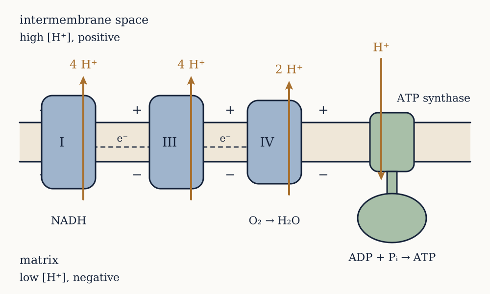
That this is the mechanism took fifteen years to accept. Through the 1950s the field looked for a "high energy intermediate", a chemical compound analogous to 1,3-bisphosphoglycerate that would be made by the respiratory chain and would then phosphorylate ADP. Dozens of laboratories searched for it and none found it.
Peter Mitchell proposed in a 1961 paper in Nature that there is no such compound, and that the intermediate is not chemical but electrical: the chain uses the energy of electron transfer to pump protons out, creating a gradient across the membrane, and ATP synthase then lets protons back in and uses the flow to make ATP. The proposal was widely rejected. It required the membrane to be intact and impermeable, made the coupling stoichiometry non-integral, and came from a scientist who had left his university post and was working in a converted manor house in Cornwall.
Two experiments settled it, and both are worth knowing because neither is a detail.
André Jagendorf and Ernest Uribe, in 1966, took chloroplast thylakoid membranes, soaked them in acid at pH 4 until the inside equilibrated, then jumped the outside to pH 8 and added ADP and phosphate. In complete darkness, with no light, no electron transfer and no substrate being oxidised, the vesicles made ATP. A pH difference alone was sufficient, which is precisely what the chemiosmotic hypothesis predicts and what no chemical intermediate theory can explain.
Efraim Racker and Walther Stoeckenius, in 1974, built the converse. They made artificial lipid vesicles containing two purified proteins with no evolutionary or functional relationship: bacteriorhodopsin, a light-driven proton pump from a salt-loving archaeon, and mitochondrial ATP synthase from cow heart. Shine light on the vesicles and they make ATP. Nothing connects the two proteins except the proton gradient in between, which is a direct demonstration that the gradient is the entire linkage. Mitchell received the Nobel Prize in 1978.
Example. Someone proposes that ATP synthase is driven by direct physical contact with the respiratory complexes. Which single feature of the Racker and Stoeckenius experiment refutes this, and what would you predict about the spacing of the two kinds of protein in a real mitochondrion?
The refutation is that the two proteins in the vesicle come from different domains of life and have never met in any organism, so no evolved interaction surface can exist between them, and yet the system works. Contact cannot be the mechanism. The prediction that follows is that in a real mitochondrion the respiratory complexes and the ATP synthase need not be adjacent, and they are not: electron microscopy of cristae shows ATP synthase concentrated in rows along the highly curved rims of the folds while the respiratory complexes sit in the flatter membrane, sometimes hundreds of nanometres away. That separation would be inexplicable on a direct-contact model and is unremarkable on a chemiosmotic one, because the gradient is a property of the whole compartment rather than of any location in it. It is also why the mechanism has a requirement no soluble pathway has: the membrane must be closed. Puncture the vesicle anywhere and ATP synthesis stops everywhere.
Now you. Mitchell's hypothesis was resisted partly because it predicted a non-integral number of ATP per NADH, whereas every pathway known at the time gave whole numbers. Why does chemiosmosis produce a fraction, and why is that a point in its favour rather than against it?
Answer
Because the two halves of the process are not chemically joined. In substrate-level phosphorylation one molecule hands one phosphate to one ADP, so the ratio is one by construction. In chemiosmosis the chain pumps some number of protons into a shared pool and the synthase withdraws some other number, and the two numbers are set by entirely separate pieces of machinery: how many protons a complex happens to translocate per electron pair, and how many subunits happen to be in the synthase's rotor ring. There is no reason for one to divide the other. That it is a point in favour becomes clear once the rotor rings are counted, because they differ between organisms: eight subunits in mammalian mitochondria, ten in yeast, up to fifteen in some bacteria, which means the protons per ATP differ between species running otherwise identical chemistry. A chemical intermediate model cannot accommodate that at all, while a gradient model predicts exactly this kind of variation, and predicts further that an organism living on a shallow gradient should have a larger ring. Larger rings are indeed found in organisms with weak proton-motive force.
The size of the gradient
The proton-motive force has two parts, because a proton carries both concentration and charge:
In a working mitochondrion the membrane potential is about 160 mV, inside negative, and the matrix is roughly 0.75 pH units more alkaline than the intermembrane space, worth about 45 mV at 37 degrees Celsius. Together is around 200 mV, and it is dominated by the electrical term rather than by the pH difference, which is not the intuitive expectation.
Each proton returning through that gradient can release kJ/mol. Ten protons per NADH therefore capture kJ of the 220 kJ available, which is 88 per cent, an efficiency at the pumping stage that no engine approaches.
The machine that sells the gradient back
ATP synthase is a rotary motor, and saying so is not a metaphor. The membrane-embedded F₀ portion contains a ring of c subunits which turns as protons pass through the interface between the ring and an adjacent subunit; the central stalk, attached to the ring, rotates inside the mushroom-shaped F₁ head that projects into the matrix, and the head contains three catalytic sites.
Paul Boyer proposed in the 1970s that the sites do not make ATP by pushing the reaction energetically, but by binding change: at any moment the three sites are in different conformations, one loosely binding ADP and phosphate, one tightly bound with ATP already formed, and one open. The remarkable claim, since confirmed, is that forming ATP from ADP and phosphate in the tight site is close to energy-neutral, because the enzyme binds ATP so much more tightly than the substrates that it pays the cost of the bond. The energy from the proton gradient is spent almost entirely on releasing the finished ATP, by rotating the stalk and forcing the site open.
John Walker's crystal structure in 1994 showed the three sites in exactly the three predicted states, and Boyer and Walker shared a Nobel Prize in 1997. Hiroyuki Noji and colleagues then made rotation visible in the same year, by fixing an F₁ head to a glass slide, attaching a fluorescently labelled actin filament to the central stalk, adding ATP, and watching the filament turn under a microscope in discrete 120-degree steps.
The stoichiometry follows from the geometry. One full rotation makes three ATP, one for each catalytic site, and requires as many protons as there are c subunits in the ring. Mammalian mitochondria have eight, so protons per ATP, and roughly one more proton per ATP is spent by the transporters that bring ADP and phosphate into the matrix and carry ATP out, giving about 3.7 in total. At 19.3 kJ per proton that is 71 kJ delivered to make a molecule worth 50 kJ, which is enough with margin, as it must be for the reaction to run at a useful rate.
Example. Making one ATP inside a cell costs about 50 kJ/mol. Using mV, work out the minimum number of protons the synthase must use, and then predict the rotor ring size of a bacterium that lives on a proton-motive force of only 120 mV.
Each proton delivers kJ/mol, so making one ATP needs at least protons, and since a rotor ring cannot deliver fractions per site the minimum practical figure is three. The mammalian ring of eight subunits gives 2.7 per ATP, which is just above that floor, consistent with a mitochondrion running at a healthy gradient and no more. Now repeat at 120 mV: each proton is worth kJ/mol, so an ATP needs protons, and a ring making three ATP per revolution must therefore have about subunits. That is a real prediction and it holds: alkaliphilic Bacillus species, which live at high external pH and therefore cannot build a large pH component to their gradient, have rotor rings of thirteen. A structural parameter of a motor can be predicted from the electrochemistry of the environment its owner lives in, which is about as direct a confirmation of a mechanism as biology offers.
Now you. Suppose the inner membrane of a mitochondrion becomes leaky, so the gradient collapses, but the cell still contains plenty of ATP made by glycolysis. What does ATP synthase do, and what would you expect a cell to have evolved in response?
Answer
It runs backwards. The enzyme is a reversible rotary motor and nothing about it enforces a direction; with no gradient to spend, the only remaining source of free energy is ATP hydrolysis, so the machine hydrolyses ATP and pumps protons out, driving the rotor the other way. This is not a curiosity: it is what happens in ischaemic tissue, and it means a cell deprived of oxygen destroys its own glycolytic ATP through its mitochondria unless something stops it. What has evolved in response is a small inhibitor protein, IF1, which binds the catalytic head and blocks the hydrolytic direction while leaving the synthetic direction unaffected. It is activated by the fall in matrix pH that accompanies loss of the gradient, so it switches on exactly under the conditions where reversal would occur. The general shape of the argument recurs throughout this course: any machine that couples two processes reversibly will run whichever way the free energy points, so a cell that needs one direction only has to spend a separate component on enforcing it.
What a glucose is actually worth
Now the whole account, per glucose. Substrate-level phosphorylation gives four ATP, two in glycolysis and two as GTP in the cycle. The reduced carriers are ten NADH, counting two from glycolysis, two from pyruvate dehydrogenase and six from two turns of the cycle, plus two FADH₂. The protons pumped are .
Divide by 3.7 protons per ATP and the chain yields 30 ATP, for a mechanistic total near 34. Measured ratios come out lower, at about 2.5 ATP per NADH and 1.5 per FADH₂, giving 32 in total, or 30 if the cell uses the cheaper of the two shuttles that carry cytosolic NADH into the mitochondrion. The gap between 34 and 32 is real and is mostly proton leak: the inner membrane is not perfectly impermeable, and some of the gradient is discharged without passing through the synthase.
So the honest figure is "about 30", not a whole number, and its imprecision is a property of the mechanism rather than of the measurement. Older textbooks give 38, which came from assuming three ATP per NADH and two per FADH₂ before anyone had measured the rotor ring.
Compare with the previous lesson. Fermentation nets two ATP per glucose and respiration nets about thirty, a factor of fifteen, and at 50 kJ per ATP the aerobic route captures per cent of the energy in the fuel. That is the payoff for the entire apparatus of membranes, complexes and gradients.
A cross-check is available and it works. An adult at rest consumes about 250 mL of oxygen per minute, which is moles per day. Each oxygen molecule accepts four electrons, that is two NADH, worth about five ATP. So the body should make moles of ATP a day, or 41 kg. The previous lesson estimated 42 kg from an entirely independent route, dietary calories divided by energy per ATP. Two calculations sharing no inputs landing within a kilogram is the sort of agreement that should raise confidence in both.
Example. 2,4-dinitrophenol is a weak acid that is lipid-soluble in both its protonated and its deprotonated form. Predict its effect on a mitochondrion, and on a person.
Being lipid soluble in both forms, it can pick up a proton on the acidic side of the membrane, diffuse across as the neutral molecule, release the proton on the alkaline side, and diffuse back as the anion. It is a proton shuttle, and it short-circuits the gradient. Electron transport continues, because oxygen is still available and the chain is not blocked, but the protons return without passing through ATP synthase, so the energy emerges entirely as heat and ATP synthesis stops. The prediction for the organism is oxygen consumption rising sharply, body temperature rising, weight falling because fuel is being burned to no purpose, and death from hyperthermia if the dose is high. All of that is documented, because DNP was sold as a slimming drug in the United States from 1933, was taken by an estimated hundred thousand people, caused deaths and cataracts, and was banned in 1938. It is still sold illegally and still kills. The pharmacological lesson is that a drug with no therapeutic ratio is not a drug: the mechanism that produces weight loss is the mechanism that produces fatal hyperthermia, and there is no dose at which one happens without the other.
Now you. Newborn humans and hibernating mammals carry brown adipose tissue, packed with mitochondria and expressing a protein called UCP1 in the inner membrane. Infants cannot shiver. What does UCP1 almost certainly do, and what does its existence say about the argument in this lesson?
Answer
It must be a regulated proton channel that lets protons back into the matrix without passing through ATP synthase, and it is: uncoupling protein 1, activated by fatty acids and inhibited by purine nucleotides, doing deliberately what dinitrophenol does indiscriminately. Fuel is oxidised at full rate and the energy appears as heat rather than ATP, which is how an infant or a hibernator warms itself without muscle activity. What its existence says about the argument is more interesting than the physiology. If the coupling between oxidation and ATP synthesis were a chemical intermediate, a protein that made a membrane leaky to protons could not possibly abolish ATP synthesis, and evolution could not have built a thermogenic tissue by adding one channel. That a single protein, doing nothing but conducting protons, converts a mitochondrion from an ATP factory into a heater is chemiosmosis demonstrated in a living animal. Physiology is here confirming a mechanism that was established in vitro, which is the direction of evidence one always hopes for and rarely gets.
The six lessons so far have built a cell that holds itself apart from the world, moves what it needs across its boundary, and pays for all of it. Every step has been carried out by a protein: a channel with carbonyls spaced to fit a potassium ion, a kinase that excludes water from its active site, a rotor with exactly eight subunits. None of that could be arrived at by chance in the lifetime of a cell, and none of it was designed. It was inherited, as a specification, from the cell that divided to make this one. The next lesson asks where that specification is kept, and the answer took four experiments over twenty-five years to establish, because everybody expected it to be the proteins themselves.